Crystal and solution structure of the C-terminal part of the Methanocaldococcus jannaschii A1AO ATP synthase subunit E revealed by X-ray diffraction and small-angle X-ray scattering
Balakrishna, A.M., Manimekalai, M.S.S., Hunke, C., Gayen, S., Rossle, M., Jeyakanthan, J., Gruber, G.(2010) J Bioenerg Biomembr 42: 311-320
- PubMed: 20571891 Search on PubMed
- DOI: https://doi.org/10.1007/s10863-010-9298-3
- Primary Citation Related Structures:
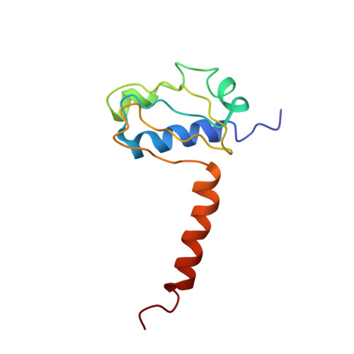
3LG8 - PubMed Abstract:
The structure of the C-terminus of subunit E (E(101-206)) of Methanocaldococcus jannaschii A-ATP synthase was determined at 4.1 A. E(101-206) consist of a N-terminal globular domain with three alpha-helices and four antiparallel beta-strands and an alpha-helix at the very C-terminus. Comparison of M. jannaschii E(101-206) with the C-terminus E(81-198) subunit E from Pyrococcus horikoshii OT3 revealed that the kink in the C-terminal alpha-helix of E(81-198), involved in dimer formation, is absent in M. jannaschii E(101-206). Whereas a major dimeric surface interface is present between the P. horikoshii E(81-198) molecules in the asymmetric unit, no such interaction could be found in the M. jannaschii E(101-206) molecules. To verify the oligomeric behaviour, the low resolution structure of the recombinant E(85-206) from M. jannaschii was determined using small angle X-ray scattering. Rigid body modeling of two copies of one of the monomer established a fit with a tail to tail arrangement.
- School of Biological Sciences, Nanyang Technological University, Singapore, Republic of Singapore.
Organizational Affiliation: