Allosteric drug discrimination is coupled to mechanochemical changes in the kinesin-5 motor core.
Kim, E.D., Buckley, R., Learman, S., Richard, J., Parke, C., Worthylake, D.K., Wojcik, E.J., Walker, R.A., Kim, S.(2010) J Biol Chem 285: 18650-18661
- PubMed: 20299460 Search on PubMedSearch on PubMed Central
- DOI: https://doi.org/10.1074/jbc.M109.092072
- Primary Citation Related Structures:
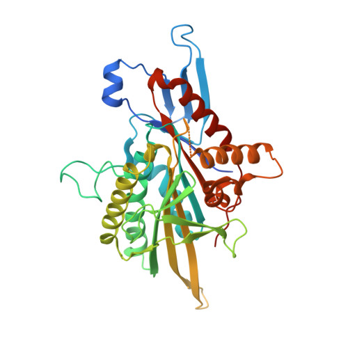
3KEN - PubMed Abstract:
Essential in mitosis, the human Kinesin-5 protein is a target for >80 classes of allosteric compounds that bind to a surface-exposed site formed by the L5 loop. Not established is why there are differing efficacies in drug inhibition. Here we compare the ligand-bound states of two L5-directed inhibitors against 15 Kinesin-5 mutants by ATPase assays and IR spectroscopy. Biochemical kinetics uncovers functional differences between individual residues at the N or C termini of the L5 loop. Infrared evaluation of solution structures and multivariate analysis of the vibrational spectra reveal that mutation and/or ligand binding not only can remodel the allosteric binding surface but also can transmit long range effects. Changes in L5-localized 3(10) helix and disordered content, regardless of substitution or drug potency, are experimentally detected. Principal component analysis couples these local structural events to two types of rearrangements in beta-sheet hydrogen bonding. These transformations in beta-sheet contacts are correlated with inhibitory drug response and are corroborated by wild type Kinesin-5 crystal structures. Despite considerable evolutionary divergence, our data directly support a theorized conserved element for long distance mechanochemical coupling in kinesin, myosin, and F(1)-ATPase. These findings also suggest that these relatively rapid IR approaches can provide structural biomarkers for clinical determination of drug sensitivity and drug efficacy in nucleotide triphosphatases.
- Department of Biochemistry and Molecular Biology, Louisiana State University Health Sciences Center, New Orleans, Louisiana 70112, USA.
Organizational Affiliation: