3K4P

Aspergillus niger Phytase


Experimental Data Snapshot

  • Method: X-RAY DIFFRACTION
  • Resolution: 2.40 Å
  • R-Value Free: 0.275 
  • R-Value Work: 0.215 
  • R-Value Observed: 0.218 

wwPDB Validation   3D Report Full Report


Ligand Structure Quality Assessment 


This is version 1.3 of the entry. See complete history


Literature

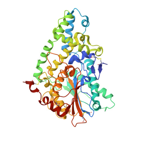
The structure of Aspergillus niger phytase PhyA in complex with a phytate mimetic

Oakley, A.J.

(2010) Biochem Biophys Res Commun 397: 745-749

  • DOI: https://doi.org/10.1016/j.bbrc.2010.06.024
  • Primary Citation of Related Structures:  
    3K4P, 3K4Q

  • PubMed Abstract: 

    Phytases hydrolyse the phosphomonoesters of phytate (myo-inositol-1,2,3,4,5,6-hexakis phosphate) and thus find uses in plant and animal production through the mobilisation of phosphorus from this source. The structure of partially deglycosylated Aspergillus niger PhyA is presented in apo form and in complex with the potent inhibitor myo-inositol-1,2,3,4,5,6-hexakis sulfate, which by analogy with phytate provides a snapshot of the Michaelis complex. The structure explains the enzyme's preference for the 3'-phosphate of phytate. The apo-and inhibitor-bound forms are similar and no induced-fit mechanism operates. Furthermore the enzyme structure is apparently unaffected by the presence of glycosides on the surface. The new structures of A. niger PhyA are discussed in the context of protein engineering studies aimed at modulating pH preference and stability.


  • Organizational Affiliation

    CSIRO Molecular and Health Technology, 343 Royal Parade, Parkville, Victoria 3052, Australia. aarono@uow.edu.au


Macromolecules
Find similar proteins by:  (by identity cutoff)  |  3D Structure
Entity ID: 1
MoleculeChains Sequence LengthOrganismDetailsImage
3-phytase A
A, B
444Aspergillus nigerMutation(s): 0 
Gene Names: phyA
EC: 3.1.3.8
UniProt
Find proteins for P34752 (Aspergillus niger)
Explore P34752 
Go to UniProtKB:  P34752
Entity Groups  
Sequence Clusters30% Identity50% Identity70% Identity90% Identity95% Identity100% Identity
UniProt GroupP34752
Sequence Annotations
Expand
  • Reference Sequence
Experimental Data & Validation

Experimental Data

  • Method: X-RAY DIFFRACTION
  • Resolution: 2.40 Å
  • R-Value Free: 0.275 
  • R-Value Work: 0.215 
  • R-Value Observed: 0.218 
  • Space Group: P 1 21 1
Unit Cell:
Length ( Å )Angle ( ˚ )
a = 71.066α = 90
b = 87.457β = 110.98
c = 82.185γ = 90
Software Package:
Software NamePurpose
Blu-Icedata collection
MOLREPphasing
REFMACrefinement
MOSFLMdata reduction
SCALAdata scaling

Structure Validation

View Full Validation Report


Ligand Structure Quality Assessment 


Entry History 

Deposition Data

  • Released Date: 2010-06-30 
  • Deposition Author(s): Oakley, A.J.

Revision History  (Full details and data files)

  • Version 1.0: 2010-06-30
    Type: Initial release
  • Version 1.1: 2011-07-13
    Changes: Version format compliance
  • Version 1.2: 2020-07-29
    Type: Remediation
    Reason: Carbohydrate remediation
    Changes: Data collection, Derived calculations, Structure summary
  • Version 1.3: 2023-11-01
    Changes: Data collection, Database references, Refinement description, Structure summary