X-ray crystallographic and NMR studies of pantothenate synthetase provide insights into the mechanism of homotropic inhibition by pantoate
Chakrabarti, K.S., Thakur, K.G., Gopal, B., Sarma, S.P.(2010) FEBS J 277: 697-712
- PubMed: 20059543 Search on PubMed
- DOI: https://doi.org/10.1111/j.1742-4658.2009.07515.x
- Primary Citation Related Structures:
3GUZ - PubMed Abstract:
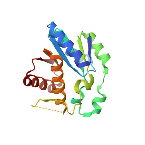
The structural basis for the homotropic inhibition of pantothenate synthetase by the substrate pantoate was investigated by X-ray crystallography and high-resolution NMR spectroscopic methods. The tertiary structure of the dimeric N-terminal domain of Escherichia coli pantothenate synthetase, determined by X-ray crystallography to a resolution of 1.7 A, showed a second molecule of pantoate bound in the ATP-binding pocket. Pantoate binding to the ATP-binding site induced large changes in structure, mainly for backbone and side chain atoms of residues in the ATP binding HXGH(34-37) motif. Sequence-specific NMR resonance assignments and solution secondary structure of the dimeric N-terminal domain, obtained using samples enriched in (2)H, (13)C, and (15)N, indicated that the secondary structural elements were conserved in solution. Nitrogen-15 edited two-dimensional solution NMR chemical shift mapping experiments revealed that pantoate, at 10 mm, bound at these two independent sites. The solution NMR studies unambiguously demonstrated that ATP stoichiometrically displaced pantoate from the ATP-binding site. All NMR and X-ray studies were conducted at substrate concentrations used for enzymatic characterization of pantothenate synthetase from different sources [Jonczyk R & Genschel U (2006) J Biol Chem 281, 37435-37446]. As pantoate binding to its canonical site is structurally conserved, these results demonstrate that the observed homotropic effects of pantoate on pantothenate biosynthesis are caused by competitive binding of this substrate to the ATP-binding site. The results presented here have implications for the design and development of potential antibacterial and herbicidal agents.
- Molecular Biophysics Unit, Indian Institute of Science, Bangalore, India.
Organizational Affiliation: