3FXR | pdb_00003fxr

Crystal structure of TsaR in complex with sulfate


Experimental Data Snapshot

  • Method: X-RAY DIFFRACTION
  • Resolution: 2.50 Å
  • R-Value Free: 
    0.269 (Depositor), 0.276 (DCC) 
  • R-Value Work: 
    0.204 (Depositor), 0.205 (DCC) 
  • R-Value Observed: 
    0.207 (Depositor) 

Starting Model: experimental
View more details

wwPDB Validation 3D Report Full Report

Validation slider image for 3FXR

Ligand Structure Quality Assessment 


This is version 1.4 of the entry. See complete history

Literature

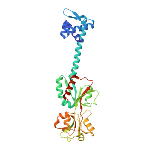
Structural studies on the full-length LysR-type regulator TsaR from Comamonas testosteroni T-2 reveal a novel open conformation of the tetrameric LTTR fold

Monferrer, D.Tralau, T.Kertesz, M.A.Dix, I.Sola, M.Uson, I.

(2010) Mol Microbiol 75: 1199-1214

  • DOI: https://doi.org/10.1111/j.1365-2958.2010.07043.x
  • Primary Citation Related Structures: 
    3FXQ, 3FXR, 3FXU, 3FZJ

  • PubMed Abstract: 

    LysR-type transcriptional regulators (LTTRs) constitute the largest family of regulators in prokaryotes. The full-length structures of the LTTR TsaR from Comamonas testosteroni T-2 and its complex with the natural inducer para-toluensulfonate have been characterized by X-ray diffraction. Both ligand-free and complexed forms reveal a dramatically different quaternary structure from that of CbnR from Ralstonia eutropha, or a putative LysR-type regulator from Pseudomonas aeruginosa, the only other determined full-length structures of tetrameric LTTRs. Although all three show a head-to-head tetrameric ring, TsaR displays an open conformation, whereas CbnR and PA01-PR present additional contacts in opposing C-terminal domains that close the ring. Such large differences may be due to a broader structural versatility than previously assumed or either, reflect the intrinsic flexibility of tetrameric LTTRs. On the grounds of the sliding dimer hypothesis of LTTR activation, we propose a structural model in which the closed structures could reflect the conformation of a ligand-free LTTR, whereas inducer binding would bring about local changes to disrupt the interface linking the two compact C-terminal domains. This could lead to a TsaR-like, open structure, where the pairs of recognition helices are closer to each other by more than 10 A.


  • Organizational Affiliation
    • IBMB-CSIC, Baldiri Reixach 15, Barcelona Science Park, 08028, Barcelona, Spain.

Macromolecule Content 

  • Total Structure Weight: 68.7 kDa 
  • Atom Count: 4,909 
  • Modeled Residue Count: 592 
  • Deposited Residue Count: 610 
  • Unique protein chains: 1

Macromolecules

Find similar proteins by:|  3D Structure
Entity ID: 1
MoleculeChains  Sequence LengthOrganismDetailsImage
LysR type regulator of tsaMBCD
A, B
305Comamonas testosteroniMutation(s): 0 
Gene Names: tsaR
UniProt
Find proteins for P94678 (Comamonas testosteroni)
Explore P94678 
Go to UniProtKB:  P94678
Entity Groups
Sequence Clusters30% Identity50% Identity70% Identity90% Identity95% Identity100% Identity
UniProt GroupP94678
Sequence Annotations
Expand
Reference Sequence

Small Molecules

Ligands 3 Unique
IDChains Name / Formula / InChI Key2D Diagram3D Interactions
ASC

Query on ASC


Download:Ideal Coordinates CCD File
K [auth A]ASCORBIC ACID
C6 H8 O6
CIWBSHSKHKDKBQ-JLAZNSOCSA-N
SO4

Query on SO4


Download:Ideal Coordinates CCD File
C [auth A]
D [auth A]
E [auth A]
F [auth A]
G [auth A]
C [auth A],
D [auth A],
E [auth A],
F [auth A],
G [auth A],
H [auth A],
I [auth A],
L [auth B],
M [auth B],
N [auth B]
SULFATE ION
O4 S
QAOWNCQODCNURD-UHFFFAOYSA-L
GOL

Query on GOL


Download:Ideal Coordinates CCD File
J [auth A],
O [auth B],
P [auth B]
GLYCEROL
C3 H8 O3
PEDCQBHIVMGVHV-UHFFFAOYSA-N

Experimental Data & Validation

Experimental Data

  • Method: X-RAY DIFFRACTION
  • Resolution: 2.50 Å
  • R-Value Free:  0.269 (Depositor), 0.276 (DCC) 
  • R-Value Work:  0.204 (Depositor), 0.205 (DCC) 
  • R-Value Observed: 0.207 (Depositor) 
Space Group: C 1 2 1
Unit Cell:
Length ( Å )Angle ( ˚ )
a = 135.11α = 90
b = 51.83β = 110.52
c = 107.99γ = 90
Software Package:
Software NamePurpose
REFMACrefinement
PDB_EXTRACTdata extraction
DNAdata collection
XDSdata reduction
XSCALEdata scaling
SHARPphasing
RESOLVEphasing

Structure Validation

View Full Validation Report


Ligand Structure Quality Assessment 


Entry History 

Deposition Data

Revision History  (Full details and data files)

  • Version 1.0: 2010-01-26
    Type: Initial release
  • Version 1.1: 2011-07-13
    Changes: Advisory, Version format compliance
  • Version 1.2: 2017-11-01
    Changes: Refinement description
  • Version 1.3: 2020-07-29
    Type: Remediation
    Reason: Carbohydrate remediation
    Changes: Data collection, Database references, Derived calculations
  • Version 1.4: 2023-11-01
    Changes: Data collection, Database references, Refinement description, Structure summary