Analysis of the structural determinants underlying discrimination between substrate and solvent in beta-phosphoglucomutase catalysis.
Dai, J., Finci, L., Zhang, C., Lahiri, S., Zhang, G., Peisach, E., Allen, K.N., Dunaway-Mariano, D.(2009) Biochemistry 48: 1984-1995
- PubMed: 19154134 Search on PubMedSearch on PubMed Central
- DOI: https://doi.org/10.1021/bi801653r
- Primary Citation Related Structures:
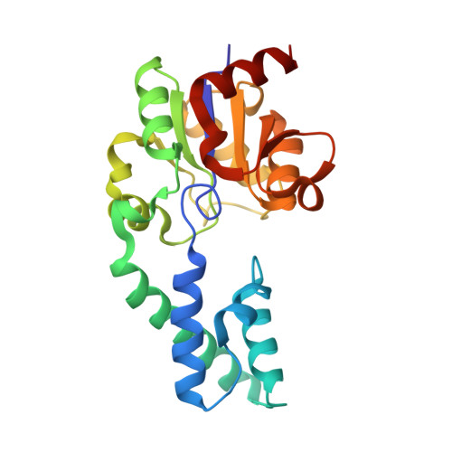
3FM9 - PubMed Abstract:
Tauhe beta-phosphoglucomutase (beta-PGM) of the haloacid dehalogenase enzyme superfamily (HADSF) catalyzes the conversion of beta-glucose 1-phosphate (betaG1P) to glucose 6-phosphate (G6P) using Asp8 of the core domain active site to mediate phosphoryl transfer from beta-glucose 1,6-(bis)phosphate (betaG1,6bisP) to betaG1P. Herein, we explore the mechanism by which hydrolysis of the beta-PGM phospho-Asp8 is avoided during the time that the active site must remain open to solvent to allow the exchange of the bound product G6P with the substrate betaG1P. On the basis of structural information, a model of catalysis is proposed in which the general acid/base (Asp10) side chain moves from a position where it forms a hydrogen bond to the Thr16-Ala17 portion of the domain-domain linker to a functional position where it forms a hydrogen bond to the substrate leaving group O and a His20-Lys76 pair of the cap domain. This repositioning of the general acid/base within the core domain active site is coordinated with substrate-induced closure of the cap domain over the core domain. The model predicts that Asp10 is required for general acid/base catalysis and for stabilization of the enzyme in the cap-closed conformation. It also predicts that hinge residue Thr16 plays a key role in productive domain-domain association, that hydrogen bond interaction with the Thr16 backbone amide NH group is required to prevent phospho-Asp8 hydrolysis in the cap-open conformation, and that the His20-Lys76 pair plays an important role in substrate-induced cap closure. The model is examined via kinetic analyses of Asp10, Thr16, His20, and Lys76 site-directed mutants. Replacement of Asp10 with Ala, Ser, Cys, Asn, or Glu resulted in no observable activity. The kinetic consequences of the replacement of linker residue Thr16 with Pro include a reduced rate of Asp8 phosphorylation by betaG1,6bisP, a reduced rate of cycling of the phosphorylated enzyme to convert betaG1P to G6P, and an enhanced rate of phosphoryl transfer from phospho-Asp8 to water. The X-ray crystal structure of the T16P mutant at 2.7 A resolution provides a snapshot of the enzyme in an unnatural cap-open conformation where the Asp10 side chain is located in the core domain active site. The His20 and Lys76 site-directed mutants exhibit reduced activity in catalysis of the Asp8-mediated phosphoryl transfer between betaG1,6bisP and betaG1P but no reduction in the rate of phospho-Asp8 hydrolysis. Taken together, the results support a substrate induced-fit model of catalysis in which betaG1P binding to the core domain facilitates recruitment of the general acid/base Asp10 to the catalytic site and induces cap closure.
- Department of Chemistry, University of New Mexico, Albuquerque, New Mexico 87131, USA.
Organizational Affiliation: