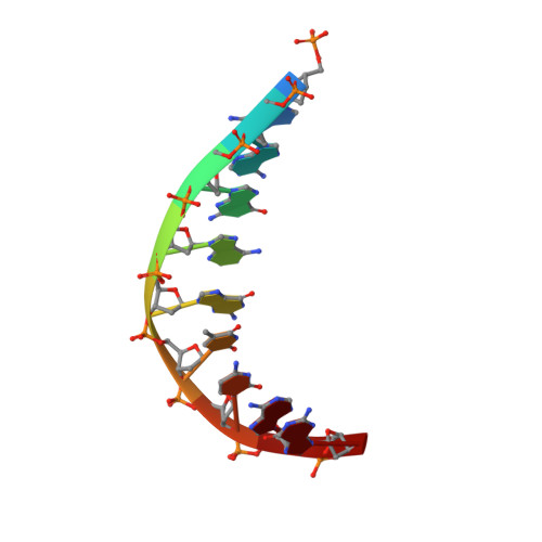
3E54

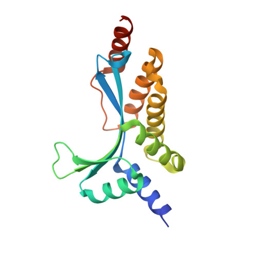
Archaeal Intron-encoded Homing Endonuclease I-Vdi141I Complexed With DNA


Experimental Data Snapshot

  • Method: X-RAY DIFFRACTION
  • Resolution: 2.50 Å
  • R-Value Free: 0.250 
  • R-Value Work: 0.231 
  • R-Value Observed: 0.231 

wwPDB Validation   3D Report Full Report


This is version 1.4 of the entry. See complete history


Literature

Recognition of a common rDNA target site in archaea and eukarya by analogous LAGLIDADG and His-Cys box homing endonucleases

Nomura, N.Nomura, Y.Sussman, D.Klein, D.Stoddard, B.L.

(2008) Nucleic Acids Res 36: 6988-6998

  • DOI: https://doi.org/10.1093/nar/gkn846
  • Primary Citation of Related Structures:  
    3E54

  • PubMed Abstract: 

    The presence of a homing endonuclease gene (HEG) within a microbial intron or intein empowers the entire element with the ability to invade genomic targets. The persistence of a homing endonuclease lineage depends in part on conservation of its DNA target site. One such rDNA sequence has been invaded both in archaea and in eukarya, by LAGLIDADG and His-Cys box homing endonucleases, respectively. The bases encoded by this target include a universally conserved ribosomal structure, termed helix 69 (H69) in the large ribosomal subunit. This region forms the 'B2a' intersubunit bridge to the small ribosomal subunit, contacts bound tRNA in the A- and P-sites, and acts as a trigger for ribosome disassembly through its interactions with ribosome recycling factor. We have determined the DNA-bound structure and specificity profile of an archaeal LAGLIDADG homing endonuclease (I-Vdi141I) that recognizes this target site, and compared its specificity with the analogous eukaryal His-Cys box endonuclease I-PpoI. These homodimeric endonuclease scaffolds have arrived at similar specificity profiles across their common biological target and analogous solutions to the problem of accommodating conserved asymmetries within the DNA sequence, but with differences at individual base pairs that are fine-tuned to the sequence conservation of archaeal versus eukaryal ribosomes.


  • Organizational Affiliation

    Iwata Human Receptor Crystallography Project, ERATO, Japan Science and Technology Agency, Kyoto, Japan.


Macromolecules

Find similar proteins by:  (by identity cutoff)  |  3D Structure
Entity ID: 1
MoleculeChains Sequence LengthOrganismDetailsImage
RRNA intron-encoded endonuclease
A, B
169Vulcanisaeta distributaMutation(s): 2 
Gene Names: 23S rRNA intron (Vdi141.L1927)
EC: 3.1
UniProt
Find proteins for Q6L703 (Vulcanisaeta distributa)
Explore Q6L703 
Go to UniProtKB:  Q6L703
Entity Groups  
Sequence Clusters30% Identity50% Identity70% Identity90% Identity95% Identity100% Identity
UniProt GroupQ6L703
Sequence Annotations
Expand
  • Reference Sequence

Find similar nucleic acids by:  Sequence   |   3D Structure  

Entity ID: 2
MoleculeChains LengthOrganismImage
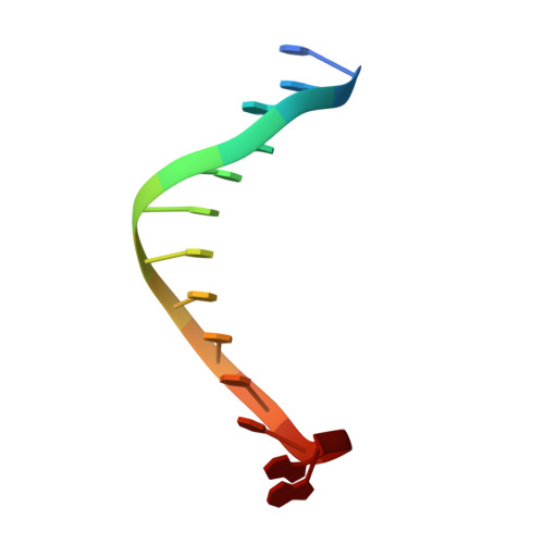
DNA (5'-D(*DCP*DTP*DGP*DAP*DCP*DTP*DCP*DTP*DCP*DTP*DTP*DAP*DA)-3')13N/A
Sequence Annotations
Expand
  • Reference Sequence

Find similar nucleic acids by:  Sequence   |   3D Structure  

Entity ID: 3
MoleculeChains LengthOrganismImage
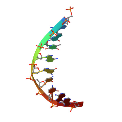
DNA (5'-D(P*DGP*DGP*DTP*DAP*DGP*DCP*DCP*DAP*DA)-3')9N/A
Sequence Annotations
Expand
  • Reference Sequence

Find similar nucleic acids by:  Sequence   |   3D Structure  

Entity ID: 4
MoleculeChains LengthOrganismImage
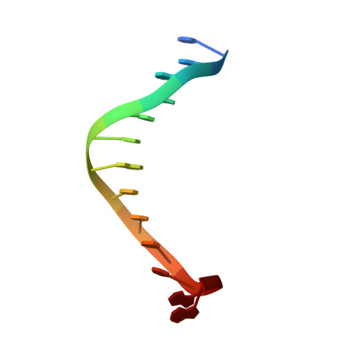
DNA (5'-D(*DTP*DTP*DGP*DGP*DCP*DTP*DAP*DCP*DCP*DTP*DTP*DAP*DA)-3')13N/A
Sequence Annotations
Expand
  • Reference Sequence

Find similar nucleic acids by:  Sequence   |   3D Structure  

Entity ID: 5
MoleculeChains LengthOrganismImage
DNA (5'-D(P*DGP*DAP*DGP*DAP*DGP*DTP*DCP*DAP*DG)-3')9N/A
Sequence Annotations
Expand
  • Reference Sequence
Small Molecules
Ligands 2 Unique
IDChains Name / Formula / InChI Key2D Diagram3D Interactions
EOH
Query on EOH

Download Ideal Coordinates CCD File 
L [auth D],
M [auth E],
N [auth F]
ETHANOL
C2 H6 O
LFQSCWFLJHTTHZ-UHFFFAOYSA-N
MG
Query on MG

Download Ideal Coordinates CCD File 
G [auth A],
H [auth A],
I [auth B],
J [auth B],
K [auth C]
MAGNESIUM ION
Mg
JLVVSXFLKOJNIY-UHFFFAOYSA-N
Experimental Data & Validation

Experimental Data

  • Method: X-RAY DIFFRACTION
  • Resolution: 2.50 Å
  • R-Value Free: 0.250 
  • R-Value Work: 0.231 
  • R-Value Observed: 0.231 
  • Space Group: P 61
Unit Cell:
Length ( Å )Angle ( ˚ )
a = 66.58α = 90
b = 66.58β = 90
c = 217.91γ = 120
Software Package:
Software NamePurpose
CNSrefinement
PDB_EXTRACTdata extraction
HKL-2000data reduction
SCALEPACKdata scaling
SOLVEphasing

Structure Validation

View Full Validation Report


Entry History 

Deposition Data

Revision History  (Full details and data files)

  • Version 1.0: 2008-12-30
    Type: Initial release
  • Version 1.1: 2011-07-13
    Changes: Version format compliance
  • Version 1.2: 2014-03-12
    Changes: Derived calculations
  • Version 1.3: 2021-11-10
    Changes: Database references, Derived calculations
  • Version 1.4: 2024-05-29
    Changes: Data collection