3DC1 | pdb_00003dc1

Crystal structure of kynurenine aminotransferase II complex with alpha-ketoglutarate


Experimental Data Snapshot

  • Method: X-RAY DIFFRACTION
  • Resolution: 2.50 Å
  • R-Value Free: 
    0.244 (Depositor), 0.242 (DCC) 
  • R-Value Work: 
    0.215 (Depositor), 0.215 (DCC) 
  • R-Value Observed: 
    0.217 (Depositor) 

Starting Model: experimental
View more details

wwPDB Validation 3D Report Full Report

Validation slider image for 3DC1

This is version 1.3 of the entry. See complete history

Literature

Substrate specificity and structure of human aminoadipate aminotransferase/kynurenine aminotransferase II

Han, Q.Cai, T.Tagle, D.A.Robinson, H.Li, J.

(2008) Biosci Rep 28: 205-215

  • DOI: https://doi.org/10.1042/BSR20080085
  • Primary Citation Related Structures: 
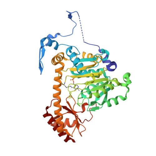
    3DC1

  • PubMed Abstract: 

    KAT (kynurenine aminotransferase) II is a primary enzyme in the brain for catalysing the transamination of kynurenine to KYNA (kynurenic acid). KYNA is the only known endogenous antagonist of the N-methyl-D-aspartate receptor. The enzyme also catalyses the transamination of aminoadipate to alpha-oxoadipate; therefore it was initially named AADAT (aminoadipate aminotransferase). As an endotoxin, aminoadipate influences various elements of glutamatergic neurotransmission and kills primary astrocytes in the brain. A number of studies dealing with the biochemical and functional characteristics of this enzyme exist in the literature, but a systematic assessment of KAT II addressing its substrate profile and kinetic properties has not been performed. The present study examines the biochemical and structural characterization of a human KAT II/AADAT. Substrate screening of human KAT II revealed that the enzyme has a very broad substrate specificity, is capable of catalysing the transamination of 16 out of 24 tested amino acids and could utilize all 16 tested alpha-oxo acids as amino-group acceptors. Kinetic analysis of human KAT II demonstrated its catalytic efficiency for individual amino-group donors and acceptors, providing information as to its preferred substrate affinity. Structural analysis of the human KAT II complex with alpha-oxoglutaric acid revealed a conformational change of an N-terminal fraction, residues 15-33, that is able to adapt to different substrate sizes, which provides a structural basis for its broad substrate specificity.


  • Organizational Affiliation
    • Department of Biochemistry, Virginia Tech, Blacksburg, VA 24061, U.S.A. qianhan@vt.edu

Macromolecule Content 

  • Total Structure Weight: 191.21 kDa 
  • Atom Count: 14,067 
  • Modeled Residue Count: 1,675 
  • Deposited Residue Count: 1,700 
  • Unique protein chains: 1

Macromolecules

Find similar proteins by:|  3D Structure
Entity ID: 1
MoleculeChains  Sequence LengthOrganismDetailsImage
Kynurenine/alpha-aminoadipate aminotransferase mitochondrial
A, B, C, D
425Homo sapiensMutation(s): 0 
Gene Names: AADATKAT2
EC: 2.6.1.7 (PDB Primary Data), 2.6.1.39 (PDB Primary Data), 2.6.1.4 (UniProt), 2.6.1.63 (UniProt), 2.6.1.73 (UniProt)
UniProt & NIH Common Fund Data Resources
Find proteins for Q8N5Z0 (Homo sapiens)
Explore Q8N5Z0 
Go to UniProtKB:  Q8N5Z0
PHAROS:  Q8N5Z0
GTEx:  ENSG00000109576 
Entity Groups
Sequence Clusters30% Identity50% Identity70% Identity90% Identity95% Identity100% Identity
UniProt GroupQ8N5Z0
Sequence Annotations
Expand
Reference Sequence

Small Molecules

Modified Residues  1 Unique
IDChains TypeFormula2D DiagramParent
LLP
Query on LLP
A, B, C, D
L-PEPTIDE LINKINGC14 H22 N3 O7 PLYS

Experimental Data & Validation

Experimental Data

  • Method: X-RAY DIFFRACTION
  • Resolution: 2.50 Å
  • R-Value Free:  0.244 (Depositor), 0.242 (DCC) 
  • R-Value Work:  0.215 (Depositor), 0.215 (DCC) 
  • R-Value Observed: 0.217 (Depositor) 
Space Group: P 1
Unit Cell:
Length ( Å )Angle ( ˚ )
a = 60.93α = 89.97
b = 72.09β = 100.65
c = 109.079γ = 93.88
Software Package:
Software NamePurpose
REFMACrefinement
HKL-2000data collection
HKL-2000data reduction
HKL-2000data scaling
PHASESphasing

Structure Validation

View Full Validation Report


Entry History 

Deposition Data

Revision History  (Full details and data files)

  • Version 1.0: 2008-07-29
    Type: Initial release
  • Version 1.1: 2011-07-13
    Changes: Non-polymer description, Version format compliance
  • Version 1.2: 2023-11-01
    Changes: Data collection, Database references, Derived calculations, Refinement description
  • Version 1.3: 2023-11-15
    Changes: Data collection