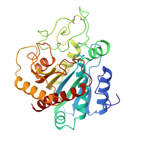
X-ray crystallographic investigation of substrate binding to carboxypeptidase A at subzero temperature.
Christianson, D.W., Lipscomb, W.N.(1986) Proc Natl Acad Sci U S A 83: 7568-7572
- PubMed: 3463986
- DOI: https://doi.org/10.1073/pnas.83.20.7568
- Primary Citation of Related Structures:
3CPA - PubMed Abstract:
A high-resolution x-ray crystallographic investigation of the complex between carboxypeptidase A (CPA; peptidyl-L-amino-acid hydrolase, EC 3.4.17.1) and the slowly hydrolyzed substrate glycyl-L-tyrosine was done at -9 degrees C. Although this enzyme-substrate complex has been the subject of earlier crystallographic investigation, a higher resolution electron-density map of the complex with greater occupancy of the substrate was desired. All crystal chemistry (i.e., crystal soaking and x-ray data collection) was performed on a diffractometer-mounted flow cell, in which the crystal was immobilized. The x-ray data to 1.6-A resolution have yielded a well-resolved structure in which the zinc ion of the active site is five-coordinate: three enzyme residues (glutamate-72, histidine-69, and histidine-196) and the carbonyl oxygen and amino terminus of glycyl-L-tyrosine complete the coordination polyhedron of the metal. These results confirm that this substrate may be bound in a nonproductive manner, because the hydrolytically important zinc-bound water has been displaced and excluded from the active site. It is likely that all dipeptide substrates of carboxypeptidase A that carry an unprotected amino terminus are poor substrates because of such favorable bidentate coordination to the metal ion of the active site.


