3CKJ

Crystal Structure of a Mycobacterial Protein


Experimental Data Snapshot

  • Method: X-RAY DIFFRACTION
  • Resolution: 1.80 Å
  • R-Value Free: 0.201 
  • R-Value Work: 0.190 
  • R-Value Observed: 0.190 

wwPDB Validation   3D Report Full Report


This is version 1.2 of the entry. See complete history


Literature

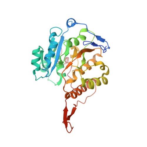
Crystal structure of a UDP-glucose-specific glycosyltransferase from a Mycobacterium species.

Fulton, Z.McAlister, A.Wilce, M.C.Brammananth, R.Zaker-Tabrizi, L.Perugini, M.A.Bottomley, S.P.Coppel, R.L.Crellin, P.K.Rossjohn, J.Beddoe, T.

(2008) J Biol Chem 283: 27881-27890

  • DOI: https://doi.org/10.1074/jbc.M801853200
  • Primary Citation of Related Structures:  
    3CKJ, 3CKN, 3CKO, 3CKQ, 3CKV

  • PubMed Abstract: 
  • Glycosyltransferases (GTs) are a large and ubiquitous family of enzymes that specifically transfer sugar moieties to a range of substrates. Mycobacterium tuberculosis contains a large number of GTs, many of which are implicated in cell wall synthesis, yet the majority of these GTs remain poorly characterized ...

    Glycosyltransferases (GTs) are a large and ubiquitous family of enzymes that specifically transfer sugar moieties to a range of substrates. Mycobacterium tuberculosis contains a large number of GTs, many of which are implicated in cell wall synthesis, yet the majority of these GTs remain poorly characterized. Here, we report the high resolution crystal structures of an essential GT (MAP2569c) from Mycobacterium avium subsp. paratuberculosis (a close homologue of Rv1208 from M. tuberculosis) in its apo- and ligand-bound forms. The structure adopted the GT-A fold and possessed the characteristic DXD motif that coordinated an Mn(2+) ion. Atypical of most GTs characterized to date, MAP2569c exhibited specificity toward the donor substrate, UDP-glucose. The structure of this ligated complex revealed an induced fit binding mechanism and provided a basis for this unique specificity. Collectively, the structural features suggested that MAP2569c may adopt a "retaining" enzymatic mechanism, which has implications for the classification of other GTs in this large superfamily.


    Organizational Affiliation

    Protein Crystallography Unit, Department of Biochemistry and Molecular Biology, School of Biomedical Sciences, Monash University, Clayton, Victoria 3800; Australian Research Council Centre of Excellence in Structural and Functional Microbial Genomics, Monash University, Clayton, Victoria 3800. Electronic address: travis.beddoe@med.monash.edu.au.


Macromolecules
Find similar proteins by:  (by identity cutoff)  |  3D Structure
Entity ID: 1
MoleculeChainsSequence LengthOrganismDetailsImage
Putative uncharacterized proteinA329N/AMutation(s): 0 
UniProt
Find proteins for Q73WU1 (Mycolicibacterium paratuberculosis (strain ATCC BAA-968 / K-10))
Explore Q73WU1 
Go to UniProtKB:  Q73WU1
Entity Groups  
Sequence Clusters30% Identity50% Identity70% Identity90% Identity95% Identity100% Identity
UniProt GroupQ73WU1
Protein Feature View
Expand
  • Reference Sequence
Small Molecules
Ligands 3 Unique
IDChainsName / Formula / InChI Key2D Diagram3D Interactions
CIT
Query on CIT

Download Ideal Coordinates CCD File 
C [auth A]CITRIC ACID
C6 H8 O7
KRKNYBCHXYNGOX-UHFFFAOYSA-N
 Ligand Interaction
MRD
Query on MRD

Download Ideal Coordinates CCD File 
D [auth A],
E [auth A],
F [auth A]
(4R)-2-METHYLPENTANE-2,4-DIOL
C6 H14 O2
SVTBMSDMJJWYQN-RXMQYKEDSA-N
 Ligand Interaction
PO4
Query on PO4

Download Ideal Coordinates CCD File 
B [auth A]PHOSPHATE ION
O4 P
NBIIXXVUZAFLBC-UHFFFAOYSA-K
 Ligand Interaction
Experimental Data & Validation

Experimental Data

  • Method: X-RAY DIFFRACTION
  • Resolution: 1.80 Å
  • R-Value Free: 0.201 
  • R-Value Work: 0.190 
  • R-Value Observed: 0.190 
  • Space Group: P 41 21 2
Unit Cell:
Length ( Å )Angle ( ˚ )
a = 86.6α = 90
b = 86.6β = 90
c = 104.3γ = 90
Software Package:
Software NamePurpose
REFMACrefinement
HKL-2000data collection
HKL-2000data reduction
HKL-2000data scaling
SnBphasing

Structure Validation

View Full Validation Report


Entry History 

Deposition Data

Revision History  (Full details and data files)

  • Version 1.0: 2008-07-29
    Type: Initial release
  • Version 1.1: 2011-07-13
    Changes: Advisory, Version format compliance
  • Version 1.2: 2017-10-25
    Changes: Refinement description