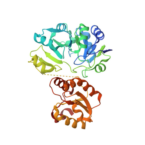
3AJE

Crystal structure of S. tokodaii Sua5 complexed with L-threonine and AMPPNP


Experimental Data Snapshot

  • Method: X-RAY DIFFRACTION
  • Resolution: 1.80 Å
  • R-Value Free: 0.238 
  • R-Value Work: 0.198 
  • R-Value Observed: 0.198 

wwPDB Validation   3D Report Full Report


Ligand Structure Quality Assessment 


This is version 1.3 of the entry. See complete history


Literature

Crystal structure of Sulfolobus tokodaii Sua5 complexed with L-threonine and AMPPNP

Kuratani, M.Kasai, T.Akasaka, R.Higashijima, K.Terada, T.Kigawa, T.Shinkai, A.Bessho, Y.Yokoyama, S.

(2011) Proteins 79: 2065-2075

  • DOI: https://doi.org/10.1002/prot.23026
  • Primary Citation of Related Structures:  
    3AJE

  • PubMed Abstract: 

    The hypermodified nucleoside N(6)-threonylcarbamoyladenosine resides at position 37 of tRNA molecules bearing U at position 36 and maintains translational fidelity in the three kingdoms of life. The N(6)-threonylcarbamoyl moiety is composed of L-threonine and bicarbonate, and its synthesis was genetically shown to require YrdC/Sua5. YrdC/Sua5 binds to tRNA and ATP. In this study, we analyzed the L-threonine-binding mode of Sua5 from the archaeon Sulfolobus tokodaii. Isothermal titration calorimetry measurements revealed that S. tokodaii Sua5 binds L-threonine more strongly than L-serine and glycine. The Kd values of Sua5 for L-threonine and L-serine are 9.3 μM and 2.6 mM, respectively. We determined the crystal structure of S. tokodaii Sua5, complexed with AMPPNP and L-threonine, at 1.8 Å resolution. The L-threonine is bound next to AMPPNP in the same pocket of the N-terminal domain. Thr118 and two water molecules form hydrogen bonds with AMPPNP in a unique manner for adenine-specific recognition. The carboxyl group and the side-chain hydroxyl and methyl groups of L-threonine are buried deep in the pocket, whereas the amino group faces AMPPNP. The L-threonine is located in a suitable position to react together with ATP for the synthesis of N(6)-threonylcarbamoyladenosine.


  • Organizational Affiliation

    RIKEN Systems and Structural Biology Center, Yokohama 230-0045, Japan.


Macromolecules
Find similar proteins by:  (by identity cutoff)  |  3D Structure
Entity ID: 1
MoleculeChains Sequence LengthOrganismDetailsImage
Putative uncharacterized protein ST1526352Sulfurisphaera tokodaii str. 7Mutation(s): 0 
Gene Names: ST1526
UniProt
Find proteins for Q970S6 (Sulfurisphaera tokodaii (strain DSM 16993 / JCM 10545 / NBRC 100140 / 7))
Explore Q970S6 
Go to UniProtKB:  Q970S6
Entity Groups  
Sequence Clusters30% Identity50% Identity70% Identity90% Identity95% Identity100% Identity
UniProt GroupQ970S6
Sequence Annotations
Expand
  • Reference Sequence
Small Molecules
Ligands 3 Unique
IDChains Name / Formula / InChI Key2D Diagram3D Interactions
ANP
Query on ANP

Download Ideal Coordinates CCD File 
C [auth A]PHOSPHOAMINOPHOSPHONIC ACID-ADENYLATE ESTER
C10 H17 N6 O12 P3
PVKSNHVPLWYQGJ-KQYNXXCUSA-N
THR
Query on THR

Download Ideal Coordinates CCD File 
B [auth A]THREONINE
C4 H9 N O3
AYFVYJQAPQTCCC-GBXIJSLDSA-N
NO3
Query on NO3

Download Ideal Coordinates CCD File 
D [auth A]
E [auth A]
F [auth A]
G [auth A]
H [auth A]
D [auth A],
E [auth A],
F [auth A],
G [auth A],
H [auth A],
I [auth A]
NITRATE ION
N O3
NHNBFGGVMKEFGY-UHFFFAOYSA-N
Binding Affinity Annotations 
IDSourceBinding Affinity
THR PDBBind:  3AJE Kd: 9300 (nM) from 1 assay(s)
Experimental Data & Validation

Experimental Data

  • Method: X-RAY DIFFRACTION
  • Resolution: 1.80 Å
  • R-Value Free: 0.238 
  • R-Value Work: 0.198 
  • R-Value Observed: 0.198 
  • Space Group: C 2 2 2
Unit Cell:
Length ( Å )Angle ( ˚ )
a = 85.35α = 90
b = 121β = 90
c = 68.01γ = 90
Software Package:
Software NamePurpose
HKL-2000data collection
MOLREPphasing
CNSrefinement
HKL-2000data reduction
SCALEPACKdata scaling

Structure Validation

View Full Validation Report


Ligand Structure Quality Assessment 


Entry History 

Deposition Data

Revision History  (Full details and data files)

  • Version 1.0: 2011-04-13
    Type: Initial release
  • Version 1.1: 2011-07-13
    Changes: Version format compliance
  • Version 1.2: 2014-01-29
    Changes: Database references
  • Version 1.3: 2023-11-01
    Changes: Data collection, Database references, Derived calculations, Refinement description