3A06

Crystal structure of DXR from Thermooga maritia, in complex with fosmidomycin and NADPH


Experimental Data Snapshot

  • Method: X-RAY DIFFRACTION
  • Resolution: 2.00 Å
  • R-Value Free: 0.240 
  • R-Value Work: 0.207 
  • R-Value Observed: 0.209 

wwPDB Validation   3D Report Full Report


Ligand Structure Quality Assessment 


This is version 1.1 of the entry. See complete history


Literature

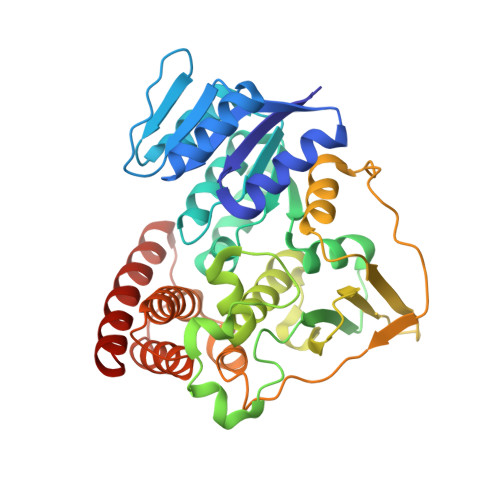
Crystal structure of 1-deoxy-d-xylulose 5-phosphate reductoisomerase from the hyperthermophile Thermotoga maritima for insights into the coordination of conformational changes and an inhibitor binding

Takenoya, M.Ohtaki, A.Noguchi, K.Endo, K.Sasaki, Y.Ohsawa, K.Yajima, S.Yohda, M.

(2010) J Struct Biol 170: 532-539

  • DOI: https://doi.org/10.1016/j.jsb.2010.03.015
  • Primary Citation of Related Structures:  
    3A06, 3A14

  • PubMed Abstract: 
  • Isopentenyl diphosphate is a precursor of various isoprenoids and is produced by the 2-C-methyl-d-erythritol 4-phosphate (MEP) pathway in plastids of plants, protozoa and many eubacteria. A key enzyme in the MEP pathway, 1-deoxy-d-xylulose 5-phosphate reductoisomerase (DXR), has been shown to be the target of fosmidomycin, which works as an antimalarial, antibacterial and herbicidal compound ...

    Isopentenyl diphosphate is a precursor of various isoprenoids and is produced by the 2-C-methyl-d-erythritol 4-phosphate (MEP) pathway in plastids of plants, protozoa and many eubacteria. A key enzyme in the MEP pathway, 1-deoxy-d-xylulose 5-phosphate reductoisomerase (DXR), has been shown to be the target of fosmidomycin, which works as an antimalarial, antibacterial and herbicidal compound. In this paper, we report studies of kinetics and the crystal structures of the thermostable DXR from the hyperthermophile Thermotoga maritima. Unlike the mesophilic DXRs, Thermotoga DXR (tDXR) showed activity only with Mg(2+) at its growth temperature. We solved the crystal structures of tDXR with and without fosmidomycin. The structure without fosmidomycin but unexpectedly bound with 2-methyl-2,4-pentanediol (MPD), revealing a new extra space available for potential drug design. This structure adopted the closed form by rigid domain rotation but without the flexible loop over the active site, which was considered as a novel conformation. Further, the conserved Asp residue responsible for cation binding seemed to play an important role in adjusting the position of fosmidomycin. Taken together, our kinetic and the crystal structures illustrate the binding mode of fosmidomycin that leads to its slow, tight binding according to the conformational changes of DXR.


    Organizational Affiliation

    Department of Biotechnology and Life Science, Graduate School of Technology, Tokyo University of Agriculture and Technology, Koganei, Tokyo 184-8588, Japan.


Macromolecules
Find similar proteins by:  (by identity cutoff)  |  3D Structure
Entity ID: 1
MoleculeChainsSequence LengthOrganismDetailsImage
1-deoxy-D-xylulose 5-phosphate reductoisomeraseA, B376Thermotoga maritimaMutation(s): 0 
Gene Names: dxr
EC: 1.1.1.267
UniProt
Find proteins for Q9WZZ1 (Thermotoga maritima (strain ATCC 43589 / DSM 3109 / JCM 10099 / NBRC 100826 / MSB8))
Explore Q9WZZ1 
Go to UniProtKB:  Q9WZZ1
Entity Groups  
Sequence Clusters30% Identity50% Identity70% Identity90% Identity95% Identity100% Identity
UniProt GroupQ9WZZ1
Protein Feature View
Expand
  • Reference Sequence
Small Molecules
Ligands 3 Unique
IDChainsName / Formula / InChI Key2D Diagram3D Interactions
NDP
Query on NDP

Download Ideal Coordinates CCD File 
C [auth A],
D [auth A],
G [auth B]
NADPH DIHYDRO-NICOTINAMIDE-ADENINE-DINUCLEOTIDE PHOSPHATE
C21 H30 N7 O17 P3
ACFIXJIJDZMPPO-NNYOXOHSSA-N
 Ligand Interaction
FOM
Query on FOM

Download Ideal Coordinates CCD File 
E [auth A],
H [auth B]
3-[FORMYL(HYDROXY)AMINO]PROPYLPHOSPHONIC ACID
C4 H10 N O5 P
GJXWDTUCERCKIX-UHFFFAOYSA-N
 Ligand Interaction
MG
Query on MG

Download Ideal Coordinates CCD File 
F [auth A],
I [auth B]
MAGNESIUM ION
Mg
JLVVSXFLKOJNIY-UHFFFAOYSA-N
 Ligand Interaction
Binding Affinity Annotations 
IDSourceBinding Affinity
FOM Binding MOAD:  3A06 IC50: 100 (nM) from 1 assay(s)
Experimental Data & Validation

Experimental Data

  • Method: X-RAY DIFFRACTION
  • Resolution: 2.00 Å
  • R-Value Free: 0.240 
  • R-Value Work: 0.207 
  • R-Value Observed: 0.209 
  • Space Group: P 43
Unit Cell:
Length ( Å )Angle ( ˚ )
a = 108.622α = 90
b = 108.622β = 90
c = 74.662γ = 90
Software Package:
Software NamePurpose
REFMACrefinement
HKL-2000data collection
HKL-2000data reduction
SCALEPACKdata scaling
MOLREPphasing

Structure Validation

View Full Validation Report


Ligand Structure Quality Assessment 


Entry History 

Deposition Data

Revision History  (Full details and data files)

  • Version 1.0: 2010-03-16
    Type: Initial release
  • Version 1.1: 2011-07-13
    Changes: Version format compliance