3ZNC

MURINE CARBONIC ANHYDRASE IV COMPLEXED WITH BRINZOLAMIDE


Experimental Data Snapshot

  • Method: X-RAY DIFFRACTION
  • Resolution: 2.80 Å
  • R-Value Free: 0.277 
  • R-Value Work: 0.191 
  • R-Value Observed: 0.191 

wwPDB Validation   3D Report Full Report


This is version 1.2 of the entry. See complete history


Literature

Structures of murine carbonic anhydrase IV and human carbonic anhydrase II complexed with brinzolamide: molecular basis of isozyme-drug discrimination.

Stams, T.Chen, Y.Boriack-Sjodin, P.A.Hurt, J.D.Liao, J.May, J.A.Dean, T.Laipis, P.Silverman, D.N.Christianson, D.W.

(1998) Protein Sci 7: 556-563

  • DOI: https://doi.org/10.1002/pro.5560070303
  • Primary Citation of Related Structures:  
    1A42, 2ZNC, 3ZNC

  • PubMed Abstract: 

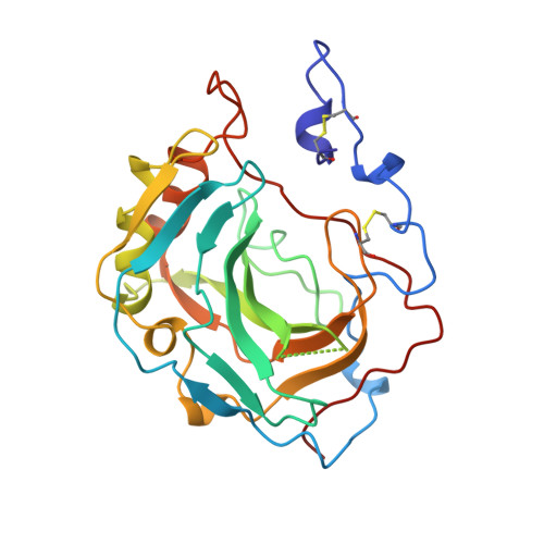
    Carbonic anhydrase IV (CAIV) is a membrane-associated enzyme anchored to plasma membrane surfaces by a phosphatidylinositol glycan linkage. We have determined the 2.8-angstroms resolution crystal structure of a truncated, soluble form of recombinant murine CAIV. We have also determined the structure of its complex with a drug used for glaucoma therapy, the sulfonamide inhibitor brinzolamide (Azopt). The overall structure of murine CAIV is generally similar to that of human CAIV; however, some local structural differences are found in the active site resulting from amino acid sequence differences in the "130's segment" and the residue-63 loop (these may affect the nearby catalytic proton shuttle, His-64). Similar to human CAIV, the C-terminus of murine CAIV is surrounded by a substantial electropositive surface potential that may stabilize the interaction with the phospholipid membrane. Binding interactions observed for brinzolamide rationalize the generally weaker affinity of inhibitors used in glaucoma therapy toward CAIV compared with CAII.


  • Organizational Affiliation

    Roy and Diana Vagelos Laboratories, Department of Chemistry, University of Pennsylvania, Philadelphia 19104-6323, USA.


Macromolecules
Find similar proteins by:  (by identity cutoff)  |  3D Structure
Entity ID: 1
MoleculeChains Sequence LengthOrganismDetailsImage
CARBONIC ANHYDRASE IV258Mus musculusMutation(s): 0 
Gene Names: MURINE CAIV
EC: 4.2.1.1
UniProt & NIH Common Fund Data Resources
Find proteins for Q64444 (Mus musculus)
Explore Q64444 
Go to UniProtKB:  Q64444
IMPC:  MGI:1096574
Entity Groups  
Sequence Clusters30% Identity50% Identity70% Identity90% Identity95% Identity100% Identity
UniProt GroupQ64444
Sequence Annotations
Expand
  • Reference Sequence
Small Molecules
Ligands 2 Unique
IDChains Name / Formula / InChI Key2D Diagram3D Interactions
BZ1
Query on BZ1

Download Ideal Coordinates CCD File 
C [auth A](+)-4-ETHYLAMINO-3,4-DIHYDRO-2-(METHOXY)PROPYL-2H-THIENO[3,2-E]-1,2-THIAZINE-6-SULFONAMIDE-1,1-DIOXIDE
C12 H21 N3 O5 S3
HCRKCZRJWPKOAR-JTQLQIEISA-N
ZN
Query on ZN

Download Ideal Coordinates CCD File 
B [auth A]ZINC ION
Zn
PTFCDOFLOPIGGS-UHFFFAOYSA-N
Binding Affinity Annotations 
IDSourceBinding Affinity
BZ1 PDBBind:  3ZNC IC50: 45.3 (nM) from 1 assay(s)
Experimental Data & Validation

Experimental Data

  • Method: X-RAY DIFFRACTION
  • Resolution: 2.80 Å
  • R-Value Free: 0.277 
  • R-Value Work: 0.191 
  • R-Value Observed: 0.191 
  • Space Group: C 2 2 21
Unit Cell:
Length ( Å )Angle ( ˚ )
a = 53.5α = 90
b = 85.2β = 90
c = 121.9γ = 90
Software Package:
Software NamePurpose
MOSFLMdata reduction
SCALAdata scaling
X-PLORmodel building
X-PLORrefinement
CCP4data scaling
X-PLORphasing

Structure Validation

View Full Validation Report


Entry History 

Deposition Data

Revision History  (Full details and data files)

  • Version 1.0: 1999-03-23
    Type: Initial release
  • Version 1.1: 2008-03-25
    Changes: Version format compliance
  • Version 1.2: 2011-07-13
    Changes: Version format compliance