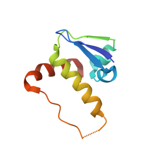
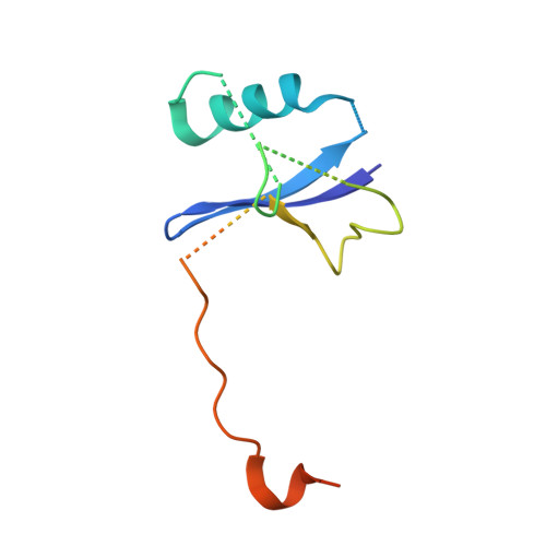
3ZKJ

Crystal Structure of Ankyrin Repeat and Socs Box-Containing Protein 9 (Asb9) in Complex with Elonginb and Elonginc


Experimental Data Snapshot

  • Method: X-RAY DIFFRACTION
  • Resolution: 2.58 Å
  • R-Value Free: 0.265 
  • R-Value Work: 0.227 
  • R-Value Observed: 0.229 

wwPDB Validation   3D Report Full Report


This is version 1.4 of the entry. See complete history


Literature

Molecular Architecture of the Ankyrin Socs Box Family of Cul5-Dependent E3 Ubiquitin Ligases

Muniz, J.R.C.Guo, K.Kershaw, N.J.Ayinampudi, V.von Delft, F.Babon, J.J.Bullock, A.N.

(2013) J Mol Biol 425: 3166

  • DOI: https://doi.org/10.1016/j.jmb.2013.06.015
  • Primary Citation of Related Structures:  
    2WZK, 3ZKJ

  • PubMed Abstract: 
  • Multi-subunit Cullin-RING E3 ligases often use repeat domain proteins as substrate-specific adaptors. Structures of these macromolecular assemblies are determined for the F-box-containing leucine-rich repeat and WD40 repeat families, but not for the suppressor of cytokine signaling (SOCS)-box-containing ankyrin repeat proteins (ASB1-18), which assemble with Elongins B and C and Cul5 ...

    Multi-subunit Cullin-RING E3 ligases often use repeat domain proteins as substrate-specific adaptors. Structures of these macromolecular assemblies are determined for the F-box-containing leucine-rich repeat and WD40 repeat families, but not for the suppressor of cytokine signaling (SOCS)-box-containing ankyrin repeat proteins (ASB1-18), which assemble with Elongins B and C and Cul5. We determined the crystal structures of the ternary complex of ASB9-Elongin B/C as well as the interacting N-terminal domain of Cul5 and used structural comparisons to establish a model for the complete Cul5-based E3 ligase. The structures reveal a distinct architecture of the ASB9 complex that positions the ankyrin domain coaxial to the SOCS box-Elongin B/C complex and perpendicular to other repeat protein complexes. This alternative architecture appears favorable to present the ankyrin domain substrate-binding site to the E2-ubiquitin, while also providing spacing suitable for bulky ASB9 substrates, such as the creatine kinases. The presented Cul5 structure also differs from previous models and deviates from other Cullins via a rigid-body rotation between Cullin repeats. This work highlights the adaptability of repeat domain proteins as scaffolds in substrate recognition and lays the foundation for future structure-function studies of this important E3 family.


    Organizational Affiliation

    Structural Genomics Consortium, University of Oxford, Old Road Campus Research Building, Roosevelt Drive, Oxford OX3 7DQ, United Kingdom.


Macromolecules
Find similar proteins by:  (by identity cutoff)  |  3D Structure
Entity ID: 1
MoleculeChainsSequence LengthOrganismDetailsImage
ANKYRIN REPEAT AND SOCS BOX PROTEIN 9A, D261Homo sapiensMutation(s): 0 
Gene Names: ASB9
UniProt & NIH Common Fund Data Resources
Find proteins for Q96DX5 (Homo sapiens)
Explore Q96DX5 
Go to UniProtKB:  Q96DX5
PHAROS:  Q96DX5
Entity Groups  
Sequence Clusters30% Identity50% Identity70% Identity90% Identity95% Identity100% Identity
UniProt GroupQ96DX5
Protein Feature View
Expand
  • Reference Sequence
Find similar proteins by:  (by identity cutoff)  |  3D Structure
Entity ID: 2
MoleculeChainsSequence LengthOrganismDetailsImage
TRANSCRIPTION ELONGATION FACTOR B POLYPEPTIDE 1B, E96Homo sapiensMutation(s): 0 
Gene Names: ELOCTCEB1
UniProt & NIH Common Fund Data Resources
Find proteins for Q15369 (Homo sapiens)
Explore Q15369 
Go to UniProtKB:  Q15369
PHAROS:  Q15369
Entity Groups  
Sequence Clusters30% Identity50% Identity70% Identity90% Identity95% Identity100% Identity
UniProt GroupQ15369
Protein Feature View
Expand
  • Reference Sequence
Find similar proteins by:  (by identity cutoff)  |  3D Structure
Entity ID: 3
MoleculeChainsSequence LengthOrganismDetailsImage
TRANSCRIPTION ELONGATION FACTOR B POLYPEPTIDE 2C, F118Homo sapiensMutation(s): 0 
Gene Names: ELOBTCEB2
UniProt & NIH Common Fund Data Resources
Find proteins for Q15370 (Homo sapiens)
Explore Q15370 
Go to UniProtKB:  Q15370
PHAROS:  Q15370
Entity Groups  
Sequence Clusters30% Identity50% Identity70% Identity90% Identity95% Identity100% Identity
UniProt GroupQ15370
Protein Feature View
Expand
  • Reference Sequence
Small Molecules
Ligands 3 Unique
IDChainsName / Formula / InChI Key2D Diagram3D Interactions
PEG
Query on PEG

Download Ideal Coordinates CCD File 
H [auth A]DI(HYDROXYETHYL)ETHER
C4 H10 O3
MTHSVFCYNBDYFN-UHFFFAOYSA-N
 Ligand Interaction
EDO
Query on EDO

Download Ideal Coordinates CCD File 
G [auth A],
J [auth D]
1,2-ETHANEDIOL
C2 H6 O2
LYCAIKOWRPUZTN-UHFFFAOYSA-N
 Ligand Interaction
CL
Query on CL

Download Ideal Coordinates CCD File 
I [auth A],
K [auth F]
CHLORIDE ION
Cl
VEXZGXHMUGYJMC-UHFFFAOYSA-M
 Ligand Interaction
Experimental Data & Validation

Experimental Data

  • Method: X-RAY DIFFRACTION
  • Resolution: 2.58 Å
  • R-Value Free: 0.265 
  • R-Value Work: 0.227 
  • R-Value Observed: 0.229 
  • Space Group: P 21 2 21
Unit Cell:
Length ( Å )Angle ( ˚ )
a = 102.86α = 90
b = 110.74β = 90
c = 113.41γ = 90
Software Package:
Software NamePurpose
REFMACrefinement
iMOSFLMdata reduction
SCALAdata scaling
PHASERphasing

Structure Validation

View Full Validation Report


Entry History 

Revision History  (Full details and data files)

  • Version 1.0: 2013-01-30
    Type: Initial release
  • Version 1.1: 2013-07-03
    Changes: Database references, Other, Refinement description, Structure summary
  • Version 1.2: 2013-07-10
    Changes: Database references
  • Version 1.3: 2013-08-28
    Changes: Database references
  • Version 1.4: 2018-01-24
    Changes: Database references