3ZJZ | pdb_00003zjz

Open-form NavMS Sodium Channel Pore (with C-terminal Domain)


Experimental Data Snapshot

  • Method: X-RAY DIFFRACTION
  • Resolution: 2.92 Å
  • R-Value Free: 
    0.287 (Depositor), 0.285 (DCC) 
  • R-Value Work: 
    0.257 (Depositor), 0.255 (DCC) 
  • R-Value Observed: 
    0.259 (Depositor) 

Starting Model: experimental
View more details

wwPDB Validation 3D Report Full Report

Validation slider image for 3ZJZ

Ligand Structure Quality Assessment 


This is version 1.1 of the entry. See complete history

Literature

Role of the C-Terminal Domain in the Structure and Function of Tetrameric Sodium Channels.

Bagneris, C.Decaen, P.G.Hall, B.A.Naylor, C.E.Clapham, D.E.Kay, C.W.M.Wallace, B.A.

(2013) Nat Commun 4: 2465

  • DOI: https://doi.org/10.1038/ncomms3465
  • Primary Citation Related Structures: 
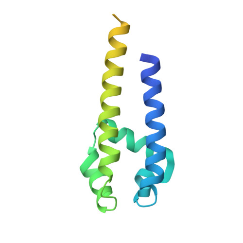
    3ZJZ

  • PubMed Abstract: 

    Voltage-gated sodium channels have essential roles in electrical signalling. Prokaryotic sodium channels are tetramers consisting of transmembrane (TM) voltage-sensing and pore domains, and a cytoplasmic carboxy-terminal domain. Previous crystal structures of bacterial sodium channels revealed the nature of their TM domains but not their C-terminal domains (CTDs). Here, using electron paramagnetic resonance (EPR) spectroscopy combined with molecular dynamics, we show that the CTD of the NavMs channel from Magnetococcus marinus includes a flexible region linking the TM domains to a four-helix coiled-coil bundle. A 2.9 Å resolution crystal structure of the NavMs pore indicates the position of the CTD, which is consistent with the EPR-derived structure. Functional analyses demonstrate that the coiled-coil domain couples inactivation with channel opening, and is enabled by negatively charged residues in the linker region. A mechanism for gating is proposed based on the structure, whereby splaying of the bottom of the pore is possible without requiring unravelling of the coiled-coil.


  • Organizational Affiliation
    • 1] School of Biological Sciences, Institute of Structural and Molecular Biology, Birkbeck College, University of London, London WC1E 7HX, UK [2].

Macromolecule Content 

  • Total Structure Weight: 70.62 kDa 
  • Atom Count: 3,189 
  • Modeled Residue Count: 366 
  • Deposited Residue Count: 596 
  • Unique protein chains: 1

Macromolecules

Find similar proteins by:|  3D Structure
Entity ID: 1
MoleculeChains  Sequence LengthOrganismDetailsImage
ION TRANSPORT PROTEIN
A, B, C, D
149Magnetococcus marinus MC-1Mutation(s): 0 
Membrane Entity: Yes 
UniProt
Find proteins for A0L5S6 (Magnetococcus marinus (strain ATCC BAA-1437 / JCM 17883 / MC-1))
Explore A0L5S6 
Go to UniProtKB:  A0L5S6
Entity Groups
Sequence Clusters30% Identity50% Identity70% Identity90% Identity95% Identity100% Identity
UniProt GroupA0L5S6
Sequence Annotations
Expand
Reference Sequence

Small Molecules

Ligands 3 Unique
IDChains Name / Formula / InChI Key2D Diagram3D Interactions
12P

Query on 12P


Download:Ideal Coordinates CCD File
F [auth A],
K [auth B],
M [auth C]
DODECAETHYLENE GLYCOL
C24 H50 O13
WRZXKWFJEFFURH-UHFFFAOYSA-N
2CV

Query on 2CV


Download:Ideal Coordinates CCD File
E [auth A],
J [auth B],
L [auth C],
P [auth D]
HEGA-10
C18 H37 N O7
ITEIKACYSCODFV-ATLSCFEFSA-N
NA

Query on NA


Download:Ideal Coordinates CCD File
G [auth A],
H [auth A],
I [auth A],
N [auth C],
O [auth C]
SODIUM ION
Na
FKNQFGJONOIPTF-UHFFFAOYSA-N

Experimental Data & Validation

Experimental Data

  • Method: X-RAY DIFFRACTION
  • Resolution: 2.92 Å
  • R-Value Free:  0.287 (Depositor), 0.285 (DCC) 
  • R-Value Work:  0.257 (Depositor), 0.255 (DCC) 
  • R-Value Observed: 0.259 (Depositor) 
Space Group: C 2 2 21
Unit Cell:
Length ( Å )Angle ( ˚ )
a = 80.15α = 90
b = 334.38β = 90
c = 80.21γ = 90
Software Package:
Software NamePurpose
BUSTERrefinement
XDSdata reduction
Aimlessdata scaling
PHASERphasing

Structure Validation

View Full Validation Report


Ligand Structure Quality Assessment 


Entry History 

Deposition Data

Revision History  (Full details and data files)

  • Version 1.0: 2013-10-02
    Type: Initial release
  • Version 1.1: 2023-12-20
    Changes: Data collection, Database references, Derived calculations, Other, Refinement description