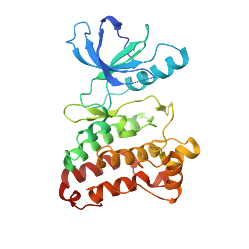
Completing the Structural Family Portrait of the Human Ephb Tyrosine Kinase Domains
Overman, R.C., Debreczeni, J.E., Truman, C.M., Mcalister, M.S., Attwood, T.K.(2014) Protein Sci 23: 627
- PubMed: 24677421
- DOI: https://doi.org/10.1002/pro.2445
- Primary Citation of Related Structures:
3ZEW, 3ZFM, 3ZFX, 3ZFY - PubMed Abstract:
The EphB receptors have key roles in cell morphology, adhesion, migration and invasion, and their aberrant action has been linked with the development and progression of many different tumor types. Their conflicting expression patterns in cancer tissues, combined with their high sequence and structural identity, present interesting challenges to those seeking to develop selective therapeutic molecules targeting this large receptor family. Here, we present the first structure of the EphB1 tyrosine kinase domain determined by X-ray crystallography to 2.5Å. Our comparative crystalisation analysis of the human EphB family kinases has also yielded new crystal forms of the human EphB2 and EphB4 catalytic domains. Unable to crystallize the wild-type EphB3 kinase domain, we used rational engineering (based on our new structures of EphB1, EphB2, and EphB4) to identify a single point mutation which facilitated its crystallization and structure determination to 2.2 Å. This mutation also improved the soluble recombinant yield of this kinase within Escherichia coli, and increased both its intrinsic stability and catalytic turnover, without affecting its ligand-binding profile. The partial ordering of the activation loop in the EphB3 structure alludes to a potential cis-phosphorylation mechanism for the EphB kinases. With the kinase domain structures of all four catalytically competent human EphB receptors now determined, a picture begins to emerge of possible opportunities to produce EphB isozyme-selective kinase inhibitors for mechanistic studies and therapeutic applications.
- AstraZeneca PLC, Alderley Park, Cheshire, SK10 4TG, United Kingdom.
Organizational Affiliation:


