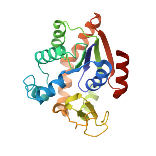
Excimer Emission Properties on Pyrene-Labeled Protein Surface: Correlation between Emission Spectra, Ring Stacking Modes, and Flexibilities of Pyrene Probes.
Fujii, A., Sekiguchi, Y., Matsumura, H., Inoue, T., Chung, W.S., Hirota, S., Matsuo, T.(2015) Bioconjug Chem 26: 537-548
- PubMed: 25646669
- DOI: https://doi.org/10.1021/acs.bioconjchem.5b00026
- Primary Citation of Related Structures:
3X2S - PubMed Abstract:
The excimer emission of pyrene is popularly employed for investigating the association between pyrene-labeled biomolecules or between pyrene-labeled places in a biomolecule. The property of pyrene excimer emission is affected by the fluctuation in ring stacking modes, which originates from the structural flexibilities of pyrene probes and/or of labeled places. Investigations of the excimer emission in terms of dynamics of pyrene stacking modes provide the detailed spatial information between pyrene-labeled places. In order to evaluate the effects of probe structures and fluctuation in pyrene-pyrene association modes on their emission properties on protein surface, three types of pyrene probe with different linker lengths were synthesized and conjugated to two cysteine residues in the A55C/C77S/V169C mutant of adenylate kinase (Adk), an enzyme that shows a structural transition between OPEN and CLOSED forms. In the CLOSED form of Adk labeled by a pyrene probe with a short linker, excimer emission was found to be predominated by the ground-state association of pyrenes. The pyrene stacking structure on the protein surface was successfully determined by an X-ray crystallographic analysis. However, the emission decay in the protein suggested the existence of several stacking orientations in solution. With the increase in the linker length, the effect of fluctuation in pyrene association modes on the spectral properties distinctly emerged at both ground and excited states. The combination of steady-state and time-resolved spectroscopic analyses is useful for differentiation in the origin of the excimer emission, which is essential for precisely understanding the interaction fashions between pyrene-labeled biomolecules.
- †Graduate School of Materials Science, Nara Institute of Science and Technology (NAIST), Ikoma, Nara 630-0192, Japan.
Organizational Affiliation:


