3WZ2 | pdb_00003wz2

Crystal structure of Pyrococcus furiosus PbaA, an archaeal homolog of proteasome-assembly chaperone


Experimental Data Snapshot

  • Method: X-RAY DIFFRACTION
  • Resolution: 2.25 Å
  • R-Value Free: 
    0.220 (Depositor), 0.218 (DCC) 
  • R-Value Work: 
    0.196 (Depositor) 
  • R-Value Observed: 
    0.198 (Depositor) 

wwPDB Validation 3D Report Full Report

Validation slider image for 3WZ2

This is version 1.3 of the entry. See complete history

Literature

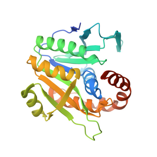
Crystal structure of archaeal homolog of proteasome-assembly chaperone PbaA

Sikdar, A.Satoh, T.Kawasaki, M.Kato, K.

(2014) Biochem Biophys Res Commun 453: 493-497

  • DOI: https://doi.org/10.1016/j.bbrc.2014.09.114
  • Primary Citation Related Structures: 
    3WZ2

  • PubMed Abstract: 

    Formation of the eukaryotic proteasome is not a spontaneous process but a highly ordered process assisted by several assembly chaperones. In contrast, archaeal proteasome subunits can spontaneously assemble into an active form. Recent bioinformatic analysis identified the proteasome-assembly chaperone-like proteins, PbaA and PbaB, in archaea. Our previous study showed that the PbaB homotetramer functions as a proteasome activator through its tentacle-like C-terminal segments. However, a functional role of the other homolog PbaA has remained elusive. Here we determined the 2.25-Å resolution structure of PbaA, illustrating its disparate tertiary and quaternary structures compared with PbaB. PbaA forms a homopentamer in which the C-terminal segments, with a putative proteasome-activating motif, are packed against the core. These findings offer deeper insights into the molecular evolution relationships between the proteasome-assembly chaperones and the proteasome activators.


  • Organizational Affiliation
    • School of Physical Science, The Graduate University for Advanced Studies, 5-1 Higashiyama, Myodaiji, Okazaki, Aichi 444-8787, Japan; Okazaki Institute for Integrative Bioscience and Institute for Molecular Science, National Institutes of Natural Sciences, 5-1 Higashiyama, Myodaiji, Okazaki, Aichi 444-8787, Japan; Graduate School of Pharmaceutical Sciences, Nagoya City University, 3-1 Tanabe-dori, Mizuho-ku, Nagoya 467-8603, Japan.

Macromolecule Content 

  • Total Structure Weight: 136.92 kDa 
  • Atom Count: 9,078 
  • Modeled Residue Count: 1,129 
  • Deposited Residue Count: 1,225 
  • Unique protein chains: 1

Macromolecules

Find similar proteins by:|  3D Structure
Entity ID: 1
MoleculeChains  Sequence LengthOrganismDetailsImage
Uncharacterized protein
A, B, C, D, E
245Pyrococcus furiosus DSM 3638Mutation(s): 0 
Gene Names: PF0015
UniProt
Find proteins for Q8U4Q9 (Pyrococcus furiosus (strain ATCC 43587 / DSM 3638 / JCM 8422 / Vc1))
Explore Q8U4Q9 
Go to UniProtKB:  Q8U4Q9
Entity Groups
Sequence Clusters30% Identity50% Identity70% Identity90% Identity95% Identity100% Identity
UniProt GroupQ8U4Q9
Sequence Annotations
Expand
Reference Sequence

Experimental Data & Validation

Experimental Data

  • Method: X-RAY DIFFRACTION
  • Resolution: 2.25 Å
  • R-Value Free:  0.220 (Depositor), 0.218 (DCC) 
  • R-Value Work:  0.196 (Depositor) 
  • R-Value Observed: 0.198 (Depositor) 
Space Group: C 2 2 21
Unit Cell:
Length ( Å )Angle ( ˚ )
a = 111.373α = 90
b = 155.187β = 90
c = 172.004γ = 90
Software Package:
Software NamePurpose
HKL-2000data collection
CRANKphasing
REFMACrefinement
DENZOdata reduction
SCALEPACKdata scaling

Structure Validation

View Full Validation Report


Entry History 

Deposition Data

Revision History  (Full details and data files)

  • Version 1.0: 2014-10-08
    Type: Initial release
  • Version 1.1: 2014-10-15
    Changes: Database references
  • Version 1.2: 2014-12-31
    Changes: Database references
  • Version 1.3: 2024-03-20
    Changes: Data collection, Database references, Refinement description