3WWC | pdb_00003wwc

The complex of pOPH_S172A of pNPB


Experimental Data Snapshot

  • Method: X-RAY DIFFRACTION
  • Resolution: 1.49 Å
  • R-Value Free: 
    0.173 (Depositor), 0.173 (DCC) 
  • R-Value Work: 
    0.154 (Depositor), 0.154 (DCC) 
  • R-Value Observed: 
    0.155 (Depositor) 

Starting Model: experimental
View more details

wwPDB Validation 3D Report Full Report

Validation slider image for 3WWC

This is version 1.2 of the entry. See complete history

Literature

Roles of tryptophan residue and disulfide bond in the variable lid region of oxidized polyvinyl alcohol hydrolase

Yang, Y.Ko, T.P.Liu, L.Li, J.Huang, C.H.Chen, J.Guo, R.T.Du, G.

(2014) Biochem Biophys Res Commun 452: 509-514

  • DOI: https://doi.org/10.1016/j.bbrc.2014.08.106
  • Primary Citation Related Structures: 
    3WWC, 3WWD, 3WWE

  • PubMed Abstract: 

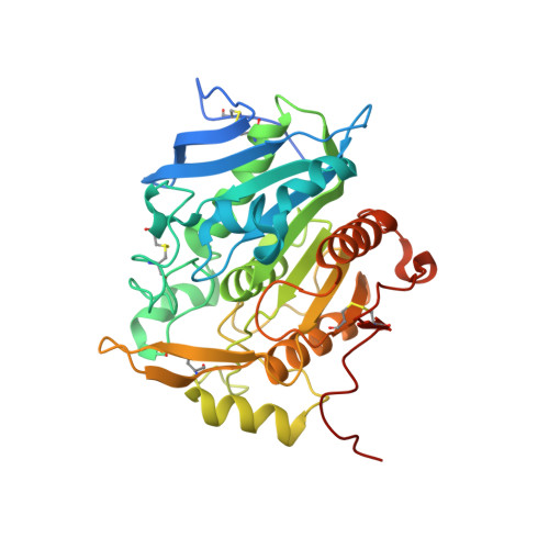
    Oxidized polyvinyl alcohol hydrolase (OPH) catalyzes the cleavage of C-C bond in β-diketone. It belongs to the α/β-hydrolase family and contains a unique lid region that covers the active site. The lid is the most variable region when pOPH from Pseudomonas sp. VM15C and sOPH from Sphingopyxis sp. 113P3 are compared. The wild-type enzymes and the pOPH mutants W255A, W255Y and W255F were analyzed for lipase activity by using p-nitrophenyl (pNP) esters as the substrates. The wild-type enzymes showed increased Km and decreased kcat/Km with the acyl chain length, and the mutants showed reduced kcat/Km for pNP acetate, indicating the importance of Trp255 in sequestering the active site from solvent. The significantly lower activity for pNP butyrate can be a result of product inhibition, as suggested by the complex crystal structures, in which butyric acid, DMSO or PEG occupied the same substrate-binding cleft. The mutant activity was retained with pNP caprylate and pNP laurate as the substrates, reflecting the amphipathic nature of the cleft. Moreover, the disulfide bond formation of Cys257/267 is important for the activity of pOPH, but it is not essential for sOPH, which has a shorter lid structure.


  • Organizational Affiliation
    • Key Laboratory of Industrial Biotechnology, Ministry of Education, Jiangnan University, Wuxi 214122, China.

Macromolecule Content 

  • Total Structure Weight: 39.62 kDa 
  • Atom Count: 3,151 
  • Modeled Residue Count: 335 
  • Deposited Residue Count: 364 
  • Unique protein chains: 1

Macromolecules

Find similar proteins by:|  3D Structure
Entity ID: 1
MoleculeChains  Sequence LengthOrganismDetailsImage
Oxidized polyvinyl alcohol hydrolase364Pseudomonas sp. VM15CMutation(s): 1 
Gene Names: pvaB
EC: 3.7.1.7
UniProt
Find proteins for Q9LCQ7 (Pseudomonas sp)
Explore Q9LCQ7 
Go to UniProtKB:  Q9LCQ7
Entity Groups
Sequence Clusters30% Identity50% Identity70% Identity90% Identity95% Identity100% Identity
UniProt GroupQ9LCQ7
Sequence Annotations
Expand
Reference Sequence

Experimental Data & Validation

Experimental Data

  • Method: X-RAY DIFFRACTION
  • Resolution: 1.49 Å
  • R-Value Free:  0.173 (Depositor), 0.173 (DCC) 
  • R-Value Work:  0.154 (Depositor), 0.154 (DCC) 
  • R-Value Observed: 0.155 (Depositor) 
Space Group: P 21 21 21
Unit Cell:
Length ( Å )Angle ( ˚ )
a = 58.326α = 90
b = 65.236β = 90
c = 84.334γ = 90
Software Package:
Software NamePurpose
Blu-Icedata collection
PHASERphasing
CNSrefinement
HKL-2000data reduction
HKL-2000data scaling

Structure Validation

View Full Validation Report


Entry History 

Deposition Data

Revision History  (Full details and data files)

  • Version 1.0: 2015-04-29
    Type: Initial release
  • Version 1.1: 2017-11-22
    Changes: Refinement description
  • Version 1.2: 2023-11-08
    Changes: Data collection, Database references, Derived calculations, Refinement description, Structure summary