Formation of domain-swapped oligomer of cytochrome C from its molten globule state oligomer.
Deshpande, M.S., Parui, P.P., Kamikubo, H., Yamanaka, M., Nagao, S., Komori, H., Kataoka, M., Higuchi, Y., Hirota, S.(2014) Biochemistry 53: 4696-4703
- PubMed: 24981551 Search on PubMed
- DOI: https://doi.org/10.1021/bi500497s
- Primary Citation Related Structures:
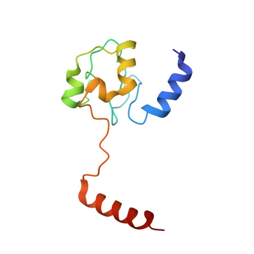
3WUI - PubMed Abstract:
Many proteins, including cytochrome c (cyt c), have been shown to form domain-swapped oligomers, but the factors governing the oligomerization process remain unrevealed. We obtained oligomers of cyt c by refolding cyt c from its acid molten globule state to neutral pH state under high protein and ion concentrations. The amount of oligomeric cyt c obtained depended on the nature of the anion (chaotropic or kosmotropic) in the solution: ClO4(-) (oligomers, 11% ± 2% (heme unit)), SCN(-) (10% ± 2%), I(-) (6% ± 2%), NO3(-) (3% ± 1%), Br(-) (2% ± 1%), Cl(-) (2% ± 1%), and SO4(2-) (3% ± 1%) for refolding of 2 mM cyt c (anion concentration 125 mM). Dimeric cyt c obtained by refolding from the molten globule state exhibited a domain-swapped structure, in which the C-terminal α-helices were exchanged between protomers. According to small-angle X-ray scattering measurements, approximately 25% of the cyt c molecules were dimerized in the molten globule state containing 125 mM ClO4(-). These results indicate that a certain amount of molten globule state oligomers of cyt c convert to domain-swapped oligomers during refolding and that the intermolecular interactions necessary for domain swapping are present in the molten globule state.
- Graduate School of Materials Science, Nara Institute of Science and Technology , 8916-5 Takayama, Ikoma, Nara 630-0192, Japan.
Organizational Affiliation: