3WU7

Spatiotemporal development of soaked protein crystal; derivative 250 sec


Experimental Data Snapshot

  • Method: X-RAY DIFFRACTION
  • Resolution: 2.00 Å
  • R-Value Free: 0.215 
  • R-Value Work: 0.167 
  • R-Value Observed: 0.169 

wwPDB Validation   3D Report Full Report


This is version 1.1 of the entry. See complete history


Literature

Spatiotemporal development of soaked protein crystal

Mizutani, R.Shimizu, Y.Saiga, R.Ueno, G.Nakamura, Y.Takeuchi, A.Uesugi, K.Suzuki, Y.

(2014) Sci Rep 4: 5731-5731

  • DOI: https://doi.org/10.1038/srep05731
  • Primary Citation of Related Structures:  
    3WPJ, 3WPK, 3WPL, 3WU7, 3WU8, 3WU9, 3WUA

  • PubMed Abstract: 

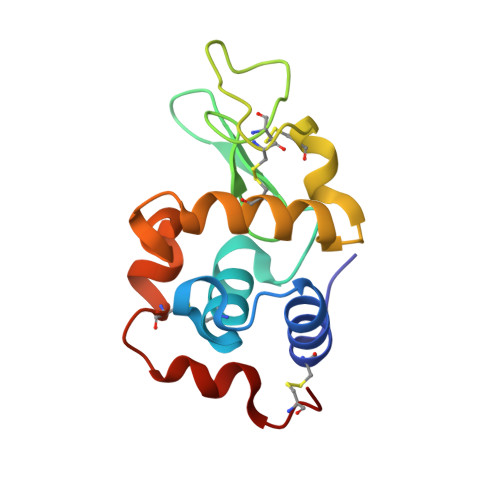
    Crystal soaking is widely performed in biological crystallography. This paper reports time-resolved X-ray crystallographic and microtomographic analyses of tetragonal crystals of chicken egg-white lysozyme soaked in mother liquor containing potassium hexachloroplatinate. The microtomographic analysis showed that X-ray attenuation spread from the superficial layer of the crystal and then to the crystal core. The crystallographic analyses indicated that platinum sites can be classified into two groups from the temporal development of the electron densities. A soaking process consisting of binding-rate-driven and equilibrium-driven layers is proposed to describe these results. This study suggests that the composition of chemical and structural species resulting from the soaking process varies depending on the position in the crystal.


  • Organizational Affiliation

    Department of Applied Biochemistry, School of Engineering, Tokai University, Kitakaname 4-1-1, Hiratsuka, Kanagawa 259-1292, Japan.


Macromolecules
Find similar proteins by:  (by identity cutoff)  |  3D Structure
Entity ID: 1
MoleculeChains Sequence LengthOrganismDetailsImage
Lysozyme C129Gallus gallusMutation(s): 0 
EC: 3.2.1.17
UniProt
Find proteins for P00698 (Gallus gallus)
Explore P00698 
Go to UniProtKB:  P00698
Entity Groups  
Sequence Clusters30% Identity50% Identity70% Identity90% Identity95% Identity100% Identity
UniProt GroupP00698
Sequence Annotations
Expand
  • Reference Sequence
Experimental Data & Validation

Experimental Data

  • Method: X-RAY DIFFRACTION
  • Resolution: 2.00 Å
  • R-Value Free: 0.215 
  • R-Value Work: 0.167 
  • R-Value Observed: 0.169 
  • Space Group: P 43 21 2
Unit Cell:
Length ( Å )Angle ( ˚ )
a = 79.174α = 90
b = 79.174β = 90
c = 37.902γ = 90
Software Package:
Software NamePurpose
BSSdata collection
REFMACrefinement
HKL-2000data reduction
SCALEPACKdata scaling

Structure Validation

View Full Validation Report


Entry History 

Deposition Data

Revision History  (Full details and data files)

  • Version 1.0: 2014-07-30
    Type: Initial release
  • Version 1.1: 2023-11-08
    Changes: Data collection, Database references, Derived calculations, Refinement description