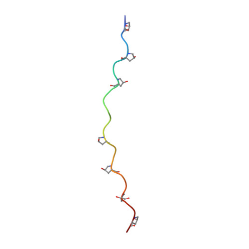
Preferred side-chain conformation of arginine residues in a triple-helical structure.
Okuyama, K., Haga, M., Noguchi, K., Tanaka, T.(2014) Biopolymers 101: 1000-1009
- PubMed: 24615532 Search on PubMed
- DOI: https://doi.org/10.1002/bip.22478
- Primary Citation Related Structures:
3WN8 - PubMed Abstract:
The crystal structure of the triple-helical peptide (Pro-Hyp-Gly)3 -Pro-Arg-Gly-(Pro-Hyp-Gly)4 (POG3-PRG-POG4) was determined at 1.45 Å resolution. POG3-PRG-POG4 was designed to permit investigation of the side-chain conformation of the Arg residues in a triple-helical structure. Because of the alternative structure of one of three Arg residues, four side-chain conformations were observed in an asymmetric unit. Among them, three adopt a ttg(-) t conformation and the other adopts a tg(-) g(-) t conformation. A statistical analysis of 80 Arg residues in various triple-helical peptides showed that, unlike those in globular proteins, they preferentially adopt a tt conformation for χ1 and χ2 , as observed in POG3-PRG-POG4. This conformation permits van der Waals contacts between the side-chain atoms of Arg and the main-chain atoms of the adjacent strand in the same molecule. Unlike many other host-guest peptides, in which there is a significant difference between the helical twists in the guest and the host peptides, POG3-PRG-POG4 shows a marked difference between the helical twists in the N-terminal peptide and those in the C-terminal peptide, separated near the Arg residue. This suggested that the unique side-chain conformation of the Arg residue affects not only the conformation of the guest peptide, but also the conformation of the peptide away from the Arg residue.
- Department of Macromolecular Science, Graduate School of Science, Osaka University, Toyonaka, Osaka, 560-0043, Japan.
Organizational Affiliation: