Crystal structure of UDP-glucose:anthocyanidin 3-O-glucosyltransferase from Clitoria ternatea
Hiromoto, T., Honjo, E., Tamada, T., Noda, N., Kazuma, K., Suzuki, M., Kuroki, R.(2013) J Synchrotron Radiat 20: 894-898
- PubMed: 24121335 Search on PubMedSearch on PubMed Central
- DOI: https://doi.org/10.1107/S0909049513020712
- Primary Citation Related Structures:
3WC4 - PubMed Abstract:
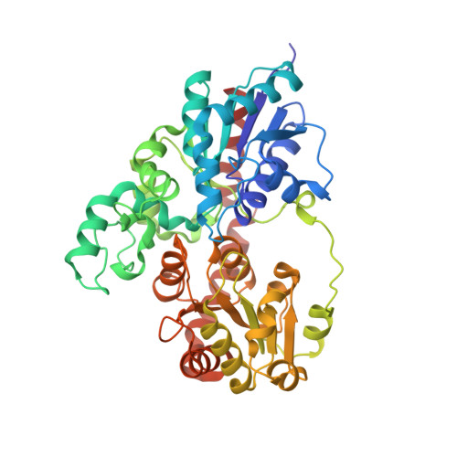
Flowers of the butterfly pea (Clitoria ternatea) accumulate a group of polyacylated anthocyanins, named ternatins, in their petals. The first step in ternatin biosynthesis is the transfer of glucose from UDP-glucose to anthocyanidins such as delphinidin, a reaction catalyzed in C. ternatea by UDP-glucose:anthocyanidin 3-O-glucosyltransferase (Ct3GT-A; AB185904). To elucidate the structure-function relationship of Ct3GT-A, recombinant Ct3GT-A was expressed in Escherichia coli and its tertiary structure was determined to 1.85 Å resolution by using X-ray crystallography. The structure of Ct3GT-A shows a common folding topology, the GT-B fold, comprised of two Rossmann-like β/α/β domains and a cleft located between the N- and C-domains containing two cavities that are used as binding sites for the donor (UDP-Glc) and acceptor substrates. By comparing the structure of Ct3GT-A with that of the flavonoid glycosyltransferase VvGT1 from red grape (Vitis vinifera) in complex with UDP-2-deoxy-2-fluoro glucose and kaempferol, locations of the catalytic His-Asp dyad and the residues involved in recognizing UDP-2-deoxy-2-fluoro glucose were essentially identical in Ct3GT-A, but certain residues of VvGT1 involved in binding kaempferol were found to be substituted in Ct3GT-A. These findings are important for understanding the differentiation of acceptor-substrate recognition in these two enzymes.
- Quantum Beam Science Directorate, Japan Atomic Energy Agency, 2-4 Shirakata-Shirane, Tokai, Ibaraki 319-1195, Japan.
Organizational Affiliation: