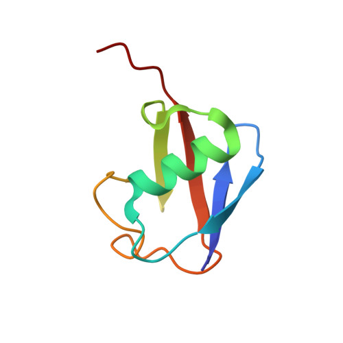
3VUY

Crystal structure of A20 ZF7 in complex with linear tetraubiquitin


Experimental Data Snapshot

  • Method: X-RAY DIFFRACTION
  • Resolution: 1.98 Å
  • R-Value Free: 0.255 
  • R-Value Work: 0.216 
  • R-Value Observed: 0.218 

wwPDB Validation   3D Report Full Report


This is version 1.0 of the entry. See complete history


Literature

Specific recognition of linear polyubiquitin by A20 zinc finger 7 is involved in NF-kappaB regulation

Tokunaga, F.Nishimasu, H.Ishitani, R.Goto, E.Noguchi, T.Mio, K.Kamei, K.Ma, A.Iwai, K.Nureki, O.

(2012) EMBO J 31: 3856-3870

  • DOI: https://doi.org/10.1038/emboj.2012.241
  • Primary Citation of Related Structures:  
    3VUW, 3VUX, 3VUY

  • PubMed Abstract: 

    LUBAC (linear ubiquitin chain assembly complex) activates the canonical NF-κB pathway through linear polyubiquitination of NEMO (NF-κB essential modulator, also known as IKKγ) and RIP1. However, the regulatory mechanism of LUBAC-mediated NF-κB activation remains elusive. Here, we show that A20 suppresses LUBAC-mediated NF-κB activation by binding linear polyubiquitin via the C-terminal seventh zinc finger (ZF7), whereas CYLD suppresses it through deubiquitinase (DUB) activity. We determined the crystal structures of A20 ZF7 in complex with linear diubiquitin at 1.70-1.98 Å resolutions. The crystal structures revealed that A20 ZF7 simultaneously recognizes the Met1-linked proximal and distal ubiquitins, and that genetic mutations associated with B cell lymphomas map to the ubiquitin-binding sites. Our functional analysis indicated that the binding of A20 ZF7 to linear polyubiquitin contributes to the recruitment of A20 into a TNF receptor (TNFR) signalling complex containing LUBAC and IκB kinase (IKK), which results in NF-κB suppression. These findings provide new insight into the regulation of immune and inflammatory responses.


  • Organizational Affiliation

    Laboratory of Molecular Cell Biology, Institute for Molecular and Cellular Regulation, Gunma University, Maebashi, Gunma, Japan. ftokunaga@gunma-u.ac.jp


Macromolecules
Find similar proteins by:  (by identity cutoff)  |  3D Structure
Entity ID: 1
MoleculeChains Sequence LengthOrganismDetailsImage
Polyubiquitin-CA,
C,
E [auth B]
76Homo sapiensMutation(s): 0 
Gene Names: UBC
UniProt & NIH Common Fund Data Resources
Find proteins for P0CG48 (Homo sapiens)
Explore P0CG48 
Go to UniProtKB:  P0CG48
PHAROS:  P0CG48
GTEx:  ENSG00000150991 
Entity Groups  
Sequence Clusters30% Identity50% Identity70% Identity90% Identity95% Identity100% Identity
UniProt GroupP0CG48
Sequence Annotations
Expand
  • Reference Sequence
Find similar proteins by:  (by identity cutoff)  |  3D Structure
Entity ID: 2
MoleculeChains Sequence LengthOrganismDetailsImage
Tumor necrosis factor alpha-induced protein 3B [auth D],
D [auth F],
F [auth E]
35Homo sapiensMutation(s): 0 
Gene Names: TNFAIP3
UniProt & NIH Common Fund Data Resources
Find proteins for P21580 (Homo sapiens)
Explore P21580 
Go to UniProtKB:  P21580
PHAROS:  P21580
GTEx:  ENSG00000118503 
Entity Groups  
Sequence Clusters30% Identity50% Identity70% Identity90% Identity95% Identity100% Identity
UniProt GroupP21580
Sequence Annotations
Expand
  • Reference Sequence
Experimental Data & Validation

Experimental Data

  • Method: X-RAY DIFFRACTION
  • Resolution: 1.98 Å
  • R-Value Free: 0.255 
  • R-Value Work: 0.216 
  • R-Value Observed: 0.218 
  • Space Group: P 31
Unit Cell:
Length ( Å )Angle ( ˚ )
a = 62.438α = 90
b = 62.438β = 90
c = 85.355γ = 120
Software Package:
Software NamePurpose
SCALEPACKdata scaling
PHENIXrefinement
PDB_EXTRACTdata extraction
BSSdata collection
HKL-2000data reduction
HKL-2000data scaling
MOLREPphasing

Structure Validation

View Full Validation Report


Entry History 

Deposition Data

Revision History  (Full details and data files)

  • Version 1.0: 2013-02-13
    Type: Initial release