Substrate recognition mechanism and substrate-dependent conformational changes of an ROK family glucokinase from Streptomyces griseus
Miyazono, K., Tabei, N., Morita, S., Ohnishi, Y., Horinouchi, S., Tanokura, M.(2012) J Bacteriol 194: 607-616
- PubMed: 22101842 Search on PubMedSearch on PubMed Central
- DOI: https://doi.org/10.1128/JB.06173-11
- Primary Citation Related Structures:
3VGK, 3VGL, 3VGM - PubMed Abstract:
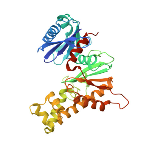
Carbon catabolite repression (CCR) is a widespread phenomenon in many bacteria that is defined as the repression of catabolic enzyme activities for an unfavorable carbon source by the presence of a preferable carbon source. In Streptomyces, secondary metabolite production often is negatively affected by the carbon source, indicating the involvement of CCR in secondary metabolism. Although the CCR mechanism in Streptomyces still is unclear, glucokinase is presumably a central player in CCR. SgGlkA, a glucokinase from S. griseus, belongs to the ROK family glucokinases, which have two consensus sequence motifs (1 and 2). Here, we report the crystal structures of apo-SgGlkA, SgGlkA in complex with glucose, and SgGlkA in complex with glucose and adenylyl imidodiphosphate (AMPPNP), which are the first structures of an ROK family glucokinase. SgGlkA is divided into a small α/β domain and a large α+β domain, and it forms a dimer-of-dimer tetrameric configuration. SgGlkA binds a β-anomer of glucose between the two domains, and His157 in consensus sequence 1 plays an important role in the glucose-binding mechanism and anomer specificity of SgGlkA. In the structures of SgGlkA, His157 forms an HC3-type zinc finger motif with three cysteine residues in consensus sequence 2 to bind a zinc ion, and it forms two hydrogen bonds with the C1 and C2 hydroxyls of glucose. When the three structures are compared, the structure of SgGlkA is found to be modified by the binding of substrates. The substrate-dependent conformational changes of SgGlkA may be related to the CCR mechanism in Streptomyces.
- Department of Applied Biological Chemistry, Graduate School of Agricultural and Life Sciences, The University of Tokyo, Bunkyo-ku, Tokyo, Japan.
Organizational Affiliation: