3VEQ | pdb_00003veq

A binary complex betwwen bovine pancreatic trypsin and a engineered mutant trypsin inhibitor


Experimental Data Snapshot

  • Method: X-RAY DIFFRACTION
  • Resolution: 2.25 Å
  • R-Value Free: 
    0.274 (Depositor), 0.273 (DCC) 
  • R-Value Work: 
    0.218 (Depositor), 0.218 (DCC) 
  • R-Value Observed: 
    0.218 (Depositor) 

Starting Model: experimental
View more details

wwPDB Validation 3D Report Full Report

Validation slider image for 3VEQ

This is version 1.3 of the entry. See complete history

Literature

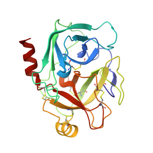
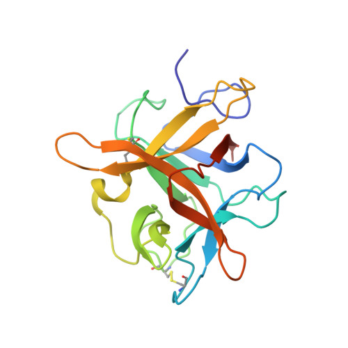
Role of remote scaffolding residues in the inhibitory loop pre-organization, flexibility, rigidification and enzyme inhibition of serine protease inhibitors

Majumder, S.Khamrui, S.Dasgupta, J.Dattagupta, J.K.Sen, U.

(2012) Biochim Biophys Acta 1824: 882-890

  • DOI: https://doi.org/10.1016/j.bbapap.2012.04.009
  • Primary Citation Related Structures: 
    3I29, 3QYD, 3VEQ

  • PubMed Abstract: 

    Canonical serine protease inhibitors interact with cognate enzymes through the P3-P2' region of the inhibitory loop while its scaffold hardly makes any contact. Neighboring scaffolding residues like Arginines or Asparagine shape-up the inhibitory loop and favor the resynthesis of cleaved scissile bond. However, role of remote scaffolding residues, which are not involved in religation, was not properly explored. Crystal structures of two engineered winged bean chymotrypsin inhibitor (WCI) complexed with Bovine trypsin (BPT) namely L65R-WCI:BPT and F64Y/L65R-WCI:BPT show that the inhibitory loop of these engineered inhibitors are recognized and rigidified properly at the enzyme active site like other strong trypsin inhibitors. Chimeric protein ETI(L)-WCI(S), having a loop of Erythrina caffra Trypsin Inhibitor, ETI on the scaffold of WCI, was previously shown to behave like substrate. Non-canonical structure of the inhibitory loop and its flexibility are attributed to the presence of smaller scaffolding residues which cannot act as barrier to the inhibitory loop like in ETI. Double mutant A76R/L115Y-(ETI(L)-WCI(S)), where the barrier is reintroduced on ETI(L)-WCI(S), shows regaining of inhibitory activity. The structure of A76R/L115Y-(ETI(L)-WCI(S)) along with L65R-WCI:BPT and F64Y/L65R-WCI:BPT demonstrate here that the lost canonical conformation of the inhibitory loop is fully restored and loop flexibility is dramatically reduced. Therefore, residues at the inhibitory loop interact with the enzyme playing the primary role in recognition and binding but scaffolding residues having no direct interaction with the enzyme are crucial for rigidification event and the inhibitory potency. B-factor analysis indicates that the amount of inhibitory loop rigidification varies between different inhibitor families.


  • Organizational Affiliation
    • Crystallography and Molecular Biology Division, Saha Institute of Nuclear Physics, Kolkata, India.

Macromolecule Content 

  • Total Structure Weight: 43.65 kDa 
  • Atom Count: 3,158 
  • Modeled Residue Count: 398 
  • Deposited Residue Count: 406 
  • Unique protein chains: 2

Macromolecules

Find similar proteins by:|  3D Structure
Entity ID: 1
MoleculeChains  Sequence LengthOrganismDetailsImage
Cationic trypsinA [auth B]223Bos taurusMutation(s): 2 
EC: 3.4.21.4
UniProt
Find proteins for P00760 (Bos taurus)
Explore P00760 
Go to UniProtKB:  P00760
Entity Groups
Sequence Clusters30% Identity50% Identity70% Identity90% Identity95% Identity100% Identity
UniProt GroupP00760
Sequence Annotations
Expand
Reference Sequence
Find similar proteins by:|  3D Structure
Entity ID: 2
MoleculeChains  Sequence LengthOrganismDetailsImage
Chymotrypsin inhibitor 3B [auth A]183Psophocarpus tetragonolobusMutation(s): 1 
UniProt
Find proteins for P10822 (Psophocarpus tetragonolobus)
Explore P10822 
Go to UniProtKB:  P10822
Entity Groups
Sequence Clusters30% Identity50% Identity70% Identity90% Identity95% Identity100% Identity
UniProt GroupP10822
Sequence Annotations
Expand
Reference Sequence

Small Molecules

Ligands 1 Unique
IDChains Name / Formula / InChI Key2D Diagram3D Interactions
CA

Query on CA


Download:Ideal Coordinates CCD File
C [auth B]CALCIUM ION
Ca
BHPQYMZQTOCNFJ-UHFFFAOYSA-N

Experimental Data & Validation

Experimental Data

  • Method: X-RAY DIFFRACTION
  • Resolution: 2.25 Å
  • R-Value Free:  0.274 (Depositor), 0.273 (DCC) 
  • R-Value Work:  0.218 (Depositor), 0.218 (DCC) 
  • R-Value Observed: 0.218 (Depositor) 
Space Group: C 1 2 1
Unit Cell:
Length ( Å )Angle ( ˚ )
a = 122.997α = 90
b = 39.826β = 119.65
c = 76.783γ = 90
Software Package:
Software NamePurpose
CNSrefinement
MAR345dtbdata collection
AUTOMARdata reduction
MOLREPphasing

Structure Validation

View Full Validation Report


Entry History 

Deposition Data

Revision History  (Full details and data files)

  • Version 1.0: 2012-10-03
    Type: Initial release
  • Version 1.1: 2013-05-08
    Changes: Database references
  • Version 1.2: 2023-11-08
    Changes: Data collection, Database references, Derived calculations, Refinement description
  • Version 1.3: 2024-10-30
    Changes: Structure summary