3V8G | pdb_00003v8g

Crystal structure of an asymmetric trimer of a glutamate transporter homologue (GltPh)


Experimental Data Snapshot

  • Method: X-RAY DIFFRACTION
  • Resolution: 4.66 Å
  • R-Value Free: 
    0.294 (Depositor), 0.328 (DCC) 
  • R-Value Work: 
    0.255 (Depositor) 
  • R-Value Observed: 
    0.257 (Depositor) 

wwPDB Validation 3D Report Full Report

Validation slider image for 3V8G

This is version 1.2 of the entry. See complete history

Literature

Crystal structure of an asymmetric trimer of a bacterial glutamate transporter homolog.

Verdon, G.Boudker, O.

(2012) Nat Struct Mol Biol 19: 355-357

  • DOI: https://doi.org/10.1038/nsmb.2233
  • Primary Citation Related Structures: 
    3V8F, 3V8G

  • PubMed Abstract: 

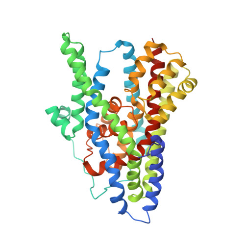
    We report a structure of a trimeric glutamate transporter homolog from Pyrococcus horikoshii with two protomers in an inward facing state and the third in an intermediate conformation between the outward and inward facing states. The intermediate shows a cavity in the thinnest region of the transporter, which is potentially accessible to extracellular and cytoplasmic solutions. Our findings suggest a structural principle by which transport intermediates may mediate uncoupled permeation of polar solutes.


  • Organizational Affiliation
    • Department of Physiology and Biophysics, Weill Cornell Medical College, New York, New York, USA.

Macromolecule Content 

  • Total Structure Weight: 268.95 kDa 
  • Atom Count: 18,038 
  • Modeled Residue Count: 2,421 
  • Deposited Residue Count: 2,532 
  • Unique protein chains: 1

Macromolecules

Find similar proteins by:|  3D Structure
Entity ID: 1
MoleculeChains  Sequence LengthOrganismDetailsImage
sodium-coupled L-aspartate transporter
A, B, C, D, E
A, B, C, D, E, F
422Pyrococcus horikoshii OT3Mutation(s): 10 
Gene Names: O59010PH1295
Membrane Entity: Yes 
UniProt
Find proteins for O59010 (Pyrococcus horikoshii (strain ATCC 700860 / DSM 12428 / JCM 9974 / NBRC 100139 / OT-3))
Explore O59010 
Go to UniProtKB:  O59010
Entity Groups
Sequence Clusters30% Identity50% Identity70% Identity90% Identity95% Identity100% Identity
UniProt GroupO59010
Sequence Annotations
Expand
Reference Sequence

Small Molecules

Ligands 2 Unique
IDChains Name / Formula / InChI Key2D Diagram3D Interactions
ASP

Query on ASP


Download:Ideal Coordinates CCD File
G [auth A]
J [auth B]
M [auth C]
P [auth D]
S [auth E]
G [auth A],
J [auth B],
M [auth C],
P [auth D],
S [auth E],
V [auth F]
ASPARTIC ACID
C4 H7 N O4
CKLJMWTZIZZHCS-REOHCLBHSA-N
NA

Query on NA


Download:Ideal Coordinates CCD File
H [auth A]
I [auth A]
K [auth B]
L [auth B]
N [auth C]
H [auth A],
I [auth A],
K [auth B],
L [auth B],
N [auth C],
O [auth C],
Q [auth D],
R [auth D],
T [auth E],
U [auth E],
W [auth F],
X [auth F]
SODIUM ION
Na
FKNQFGJONOIPTF-UHFFFAOYSA-N

Experimental Data & Validation

Experimental Data

  • Method: X-RAY DIFFRACTION
  • Resolution: 4.66 Å
  • R-Value Free:  0.294 (Depositor), 0.328 (DCC) 
  • R-Value Work:  0.255 (Depositor) 
  • R-Value Observed: 0.257 (Depositor) 
Space Group: C 1 2 1
Unit Cell:
Length ( Å )Angle ( ˚ )
a = 203.692α = 90
b = 119.627β = 113.77
c = 223.415γ = 90
Software Package:
Software NamePurpose
ADSCdata collection
PHASERphasing
REFMACrefinement
HKL-2000data reduction
HKL-2000data scaling

Structure Validation

View Full Validation Report


Entry History 

Deposition Data

Revision History  (Full details and data files)

  • Version 1.0: 2012-02-15
    Type: Initial release
  • Version 1.1: 2013-01-09
    Changes: Database references
  • Version 1.2: 2024-02-28
    Changes: Data collection, Database references, Derived calculations