3UX3

Crystal Structure of Domain-Swapped Fam96a minor dimer


Experimental Data Snapshot

  • Method: X-RAY DIFFRACTION
  • Resolution: 1.80 Å
  • R-Value Free: 0.246 
  • R-Value Work: 0.200 
  • R-Value Observed: 0.203 

Starting Model: experimental
View more details

wwPDB Validation   3D Report Full Report


This is version 1.2 of the entry. See complete history


Literature

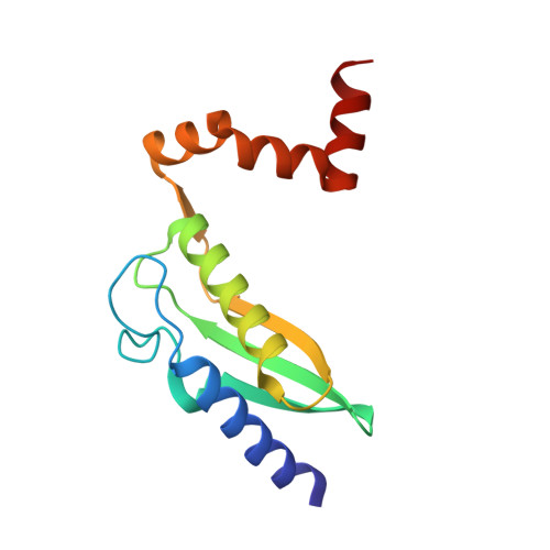
The mammalian DUF59 protein Fam96a forms two distinct types of domain-swapped dimer.

Chen, K.E.Richards, A.A.Ariffin, J.K.Ross, I.L.Sweet, M.J.Kellie, S.Kobe, B.Martin, J.L.

(2012) Acta Crystallogr D Biol Crystallogr 68: 637-648

  • DOI: https://doi.org/10.1107/S0907444912006592
  • Primary Citation of Related Structures:  
    3UX2, 3UX3

  • PubMed Abstract: 

    Fam96a mRNA, which encodes a mammalian DUF59 protein, is enriched in macrophages. Recombinant human Fam96a forms stable monomers and dimers in solution. Crystal structures of these two forms revealed that each adopts a distinct type of domain-swapped dimer, one of which is stabilized by zinc binding. Two hinge loops control Fam96a domain swapping; both are flexible and highly conserved, suggesting that domain swapping may be a common feature of eukaryotic but not bacterial DUF59 proteins. The derived monomer fold of Fam96a diverges from that of bacterial DUF59 counterparts in that the C-terminal region of Fam96a is much longer and is positioned on the opposite side of the N-terminal core fold. The putative metal-binding site of bacterial DUF59 proteins is not conserved in Fam96a, but Fam96a interacts tightly in vitro with Ciao1, the cytosolic iron-assembly protein. Moreover, Fam96a and Ciao1 can be co-immunoprecipitated, suggesting that the interaction also occurs in vivo. Although predicted to have a signal peptide, it is shown that Fam96a is cytoplasmic. The data reveal that eukaryotic DUF59 proteins share intriguing characteristics with amyloidogenic proteins.


  • Organizational Affiliation

    Institute for Molecular Bioscience, Division of Chemistry and Structural Biology, University of Queensland, Brisbane, Queensland 4072, Australia.


Macromolecules
Find similar proteins by:  (by identity cutoff)  |  3D Structure
Entity ID: 1
MoleculeChains Sequence LengthOrganismDetailsImage
MIP18 family protein FAM96A
A, B
130Homo sapiensMutation(s): 0 
Gene Names: FAM96A
UniProt & NIH Common Fund Data Resources
Find proteins for Q9H5X1 (Homo sapiens)
Explore Q9H5X1 
Go to UniProtKB:  Q9H5X1
PHAROS:  Q9H5X1
GTEx:  ENSG00000166797 
Entity Groups  
Sequence Clusters30% Identity50% Identity70% Identity90% Identity95% Identity100% Identity
UniProt GroupQ9H5X1
Sequence Annotations
Expand
  • Reference Sequence
Experimental Data & Validation

Experimental Data

  • Method: X-RAY DIFFRACTION
  • Resolution: 1.80 Å
  • R-Value Free: 0.246 
  • R-Value Work: 0.200 
  • R-Value Observed: 0.203 
  • Space Group: P 1 21 1
Unit Cell:
Length ( Å )Angle ( ˚ )
a = 49.179α = 90
b = 52.246β = 104.56
c = 65.893γ = 90
Software Package:
Software NamePurpose
SCALEPACKdata scaling
PHASERphasing
PHENIXrefinement
PDB_EXTRACTdata extraction
Blu-Icedata collection
HKL-2000data reduction
HKL-2000data scaling

Structure Validation

View Full Validation Report


Entry History 

Deposition Data

Revision History  (Full details and data files)

  • Version 1.0: 2012-05-30
    Type: Initial release
  • Version 1.1: 2020-01-01
    Changes: Advisory, Database references
  • Version 1.2: 2023-11-08
    Changes: Advisory, Data collection, Database references, Derived calculations, Refinement description