3ULR | pdb_00003ulr

Lysozyme contamination facilitates crystallization of a hetero-trimericCortactin:Arg:Lysozyme complex


Experimental Data Snapshot

  • Method: X-RAY DIFFRACTION
  • Resolution: 1.65 Å
  • R-Value Free: 
    0.225 (Depositor), 0.230 (DCC) 
  • R-Value Work: 
    0.184 (Depositor), 0.188 (DCC) 
  • R-Value Observed: 
    0.186 (Depositor) 

Starting Models: experimental
View more details

wwPDB Validation 3D Report Full Report

Validation slider image for 3ULR

This is version 1.3 of the entry. See complete history

Literature

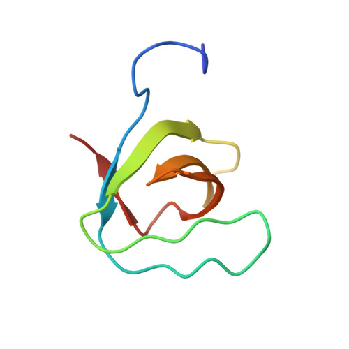
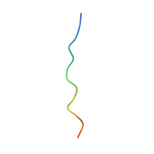
Lysozyme contamination facilitates crystallization of a heterotrimeric cortactin-Arg-lysozyme complex.

Liu, W.Macgrath, S.M.Koleske, A.J.Boggon, T.J.

(2012) Acta Crystallogr Sect F Struct Biol Cryst Commun 68: 154-158

  • DOI: https://doi.org/10.1107/S1744309111056132
  • Primary Citation Related Structures: 
    3ULR

  • PubMed Abstract: 

    Crystallization of contaminating proteins is a frequently encountered problem for macromolecular crystallographers. In this study, an attempt was made to obtain a binary cocrystal structure of the SH3 domain of cortactin and a 17-residue peptide from the Arg nonreceptor tyrosine kinase encompassing a PxxPxxPxxP (PxxP1) motif. However, cocrystals could only be obtained in the presence of trace amounts of a contaminating protein. A structure solution obtained by molecular replacement followed by ARP/wARP automatic model building allowed a 'sequence-by-crystallography' approach to discover that the contaminating protein was lysozyme. This 1.65 Å resolution crystal structure determination of a 1:1:1 heterotrimeric complex of Arg, cortactin and lysozyme thus provides an unusual `caveat emptor' warning of the dangers that underpurified proteins harbor for macromolecular crystallographers.


  • Organizational Affiliation
    • Department of Pharmacology, Yale University School of Medicine, New Haven, CT 06520, USA.

Macromolecule Content 

  • Total Structure Weight: 23.4 kDa 
  • Atom Count: 1,920 
  • Modeled Residue Count: 206 
  • Deposited Residue Count: 211 
  • Unique protein chains: 3

Macromolecules

Find similar proteins by:|  3D Structure
Entity ID: 1
MoleculeChains  Sequence LengthOrganismDetailsImage
Lysozyme C129Gallus gallusMutation(s): 0 
Gene Names: LYZ
EC: 3.2.1.17
UniProt
Find proteins for P00698 (Gallus gallus)
Explore P00698 
Go to UniProtKB:  P00698
Entity Groups
Sequence Clusters30% Identity50% Identity70% Identity90% Identity95% Identity100% Identity
UniProt GroupP00698
Sequence Annotations
Expand
Reference Sequence
Find similar proteins by:|  3D Structure
Entity ID: 2
MoleculeChains  Sequence LengthOrganismDetailsImage
Src substrate cortactin65Mus musculusMutation(s): 0 
Gene Names: CttnEms1
UniProt & NIH Common Fund Data Resources
Find proteins for Q60598 (Mus musculus)
Explore Q60598 
Go to UniProtKB:  Q60598
IMPC:  MGI:99695
Entity Groups
Sequence Clusters30% Identity50% Identity70% Identity90% Identity95% Identity100% Identity
UniProt GroupQ60598
Sequence Annotations
Expand
Reference Sequence
Find similar proteins by:  Sequence   |   3D Structure  
Entity ID: 3
MoleculeChains  Sequence LengthOrganismDetailsImage
Abelson tyrosine-protein kinase 217Homo sapiensMutation(s): 0 
EC: 2.7.10.2
UniProt & NIH Common Fund Data Resources
Find proteins for P42684 (Homo sapiens)
Explore P42684 
Go to UniProtKB:  P42684
PHAROS:  P42684
GTEx:  ENSG00000143322 
Entity Groups
Sequence Clusters30% Identity50% Identity70% Identity90% Identity95% Identity100% Identity
UniProt GroupP42684
Sequence Annotations
Expand
Reference Sequence

Experimental Data & Validation

Experimental Data

  • Method: X-RAY DIFFRACTION
  • Resolution: 1.65 Å
  • R-Value Free:  0.225 (Depositor), 0.230 (DCC) 
  • R-Value Work:  0.184 (Depositor), 0.188 (DCC) 
  • R-Value Observed: 0.186 (Depositor) 
Space Group: P 21 21 21
Unit Cell:
Length ( Å )Angle ( ˚ )
a = 38.658α = 90
b = 57.838β = 90
c = 95.513γ = 90
Software Package:
Software NamePurpose
ADSCdata collection
PHASERphasing
REFMACrefinement
HKL-2000data reduction
HKL-2000data scaling

Structure Validation

View Full Validation Report


Entry History 

Deposition Data

Revision History  (Full details and data files)

  • Version 1.0: 2012-01-11
    Type: Initial release
  • Version 1.1: 2012-02-15
    Changes: Database references
  • Version 1.2: 2023-09-13
    Changes: Data collection, Database references, Refinement description
  • Version 1.3: 2024-11-27
    Changes: Structure summary