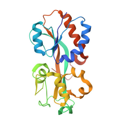
Structures of the Porphyromonas gingivalis OxyR regulatory domain explain differences in expression of the OxyR regulon in Escherichia coli and P. gingivalis.
Svintradze, D.V., Peterson, D.L., Collazo-Santiago, E.A., Lewis, J.P., Wright, H.T.(2013) Acta Crystallogr D Biol Crystallogr 69: 2091-2103
- PubMed: 24100327 Search on PubMedSearch on PubMed Central
- DOI: https://doi.org/10.1107/S0907444913019471
- Primary Citation Related Structures:
3HO7, 3T22, 3UKI - PubMed Abstract:
OxyR transcriptionally regulates Escherichia coli oxidative stress response genes through a reversibly reducible cysteine disulfide biosensor of cellular redox status. Structural changes induced by redox changes in these cysteines are conformationally transmitted to the dimer subunit interfaces, which alters dimer and tetramer interactions with DNA. In contrast to E. coli OxyR regulatory-domain structures, crystal structures of Porphyromonas gingivalis OxyR regulatory domains show minimal differences in dimer configuration on changes in cysteine disulfide redox status. This locked configuration of the P. gingivalis OxyR regulatory-domain dimer closely resembles the oxidized (activating) form of the E. coli OxyR regulatory-domain dimer. It correlates with the observed constitutive activation of some oxidative stress genes in P. gingivalis and is attributable to a single amino-acid insertion in P. gingivalis OxyR relative to E. coli OxyR. Modelling of full-length P. gingivalis, E. coli and Neisseria meningitidis OxyR-DNA complexes predicts different modes of DNA binding for the reduced and oxidized forms of each.
- OCMB Philips Institute, School of Dentistry, Virginia Commonwealth University, Richmond, VA 23298-0566, USA.
Organizational Affiliation: