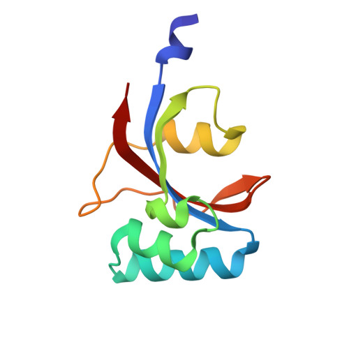
3UI5

Crystal structure of human Parvulin 14


Experimental Data Snapshot

  • Method: X-RAY DIFFRACTION
  • Resolution: 1.40 Å
  • R-Value Free: 0.137 
  • R-Value Work: 0.096 
  • R-Value Observed: 0.098 

wwPDB Validation   3D Report Full Report


Ligand Structure Quality Assessment 


This is version 1.3 of the entry. See complete history


Literature

Crystallographic proof for an extended hydrogen-bonding network in small prolyl isomerases.

Mueller, J.W.Link, N.M.Matena, A.Hoppstock, L.Ruppel, A.Bayer, P.Blankenfeldt, W.

(2011) J Am Chem Soc 133: 20096-20099

  • DOI: https://doi.org/10.1021/ja2086195
  • Primary Citation of Related Structures:  
    3UI4, 3UI5

  • PubMed Abstract: 

    Parvulins compose a family of small peptidyl-prolyl isomerases (PPIases) involved in protein folding and protein quality control. A number of amino acids in the catalytic cavity are highly conserved, but their precise role within the catalytic mechanism is unknown. The 0.8 Å crystal structure of the prolyl isomerase domain of parvulin Par14 shows the electron density of hydrogen atoms between the D74, H42, H123, and T118 side chains. This threonine residue has previously not been associated with catalysis, but a corresponding T152A mutant of Pin1 shows a dramatic reduction of catalytic activity without compromising protein stability. The observed catalytic tetrad is strikingly conserved in Pin1- and parvulin-type proteins and hence constitutes a common feature of small peptidyl prolyl isomerases.


  • Organizational Affiliation

    Institute for Structural and Medicinal Biochemistry, Center for Medical Biotechnology (ZMB), University of Duisburg-Essen, 45117 Essen, Germany.


Macromolecules
Find similar proteins by:  (by identity cutoff)  |  3D Structure
Entity ID: 1
MoleculeChains Sequence LengthOrganismDetailsImage
Peptidyl-prolyl cis-trans isomerase NIMA-interacting 4101Homo sapiensMutation(s): 0 
Gene Names: PIN4Q9Y237
EC: 5.2.1.8
UniProt & NIH Common Fund Data Resources
Find proteins for Q9Y237 (Homo sapiens)
Explore Q9Y237 
Go to UniProtKB:  Q9Y237
PHAROS:  Q9Y237
GTEx:  ENSG00000102309 
Entity Groups  
Sequence Clusters30% Identity50% Identity70% Identity90% Identity95% Identity100% Identity
UniProt GroupQ9Y237
Sequence Annotations
Expand
  • Reference Sequence
Experimental Data & Validation

Experimental Data

  • Method: X-RAY DIFFRACTION
  • Resolution: 1.40 Å
  • R-Value Free: 0.137 
  • R-Value Work: 0.096 
  • R-Value Observed: 0.098 
  • Space Group: P 21 21 21
Unit Cell:
Length ( Å )Angle ( ˚ )
a = 34.17α = 90
b = 47.06β = 90
c = 51.83γ = 90
Software Package:
Software NamePurpose
XSCALEdata scaling
REFMACrefinement
PDB_EXTRACTdata extraction

Structure Validation

View Full Validation Report


Ligand Structure Quality Assessment 


Entry History 

Deposition Data

Revision History  (Full details and data files)

  • Version 1.0: 2011-12-07
    Type: Initial release
  • Version 1.1: 2012-12-05
    Changes: Structure summary
  • Version 1.2: 2013-01-09
    Changes: Database references
  • Version 1.3: 2024-02-28
    Changes: Data collection, Database references, Derived calculations