3UEH | pdb_00003ueh

Crystal structure of human Survivin H80A mutant


Experimental Data Snapshot

  • Method: X-RAY DIFFRACTION
  • Resolution: 2.60 Å
  • R-Value Free: 
    0.224 (Depositor), 0.218 (DCC) 
  • R-Value Work: 
    0.189 (Depositor), 0.192 (DCC) 
  • R-Value Observed: 
    0.191 (Depositor) 

Starting Model: experimental
View more details

wwPDB Validation 3D Report Full Report

Validation slider image for 3UEH

This is version 1.3 of the entry. See complete history

Literature

Molecular basis for phosphospecific recognition of histone H3 tails by Survivin paralogues at inner centromeres.

Niedzialkowska, E.Wang, F.Porebski, P.J.Minor, W.Higgins, J.M.Stukenberg, P.T.

(2012) Mol Biol Cell 23: 1457-1466

  • DOI: https://doi.org/10.1091/mbc.E11-11-0904
  • Primary Citation Related Structures: 
    3UEC, 3UED, 3UEE, 3UEF, 3UEG, 3UEH, 3UEI

  • PubMed Abstract: 

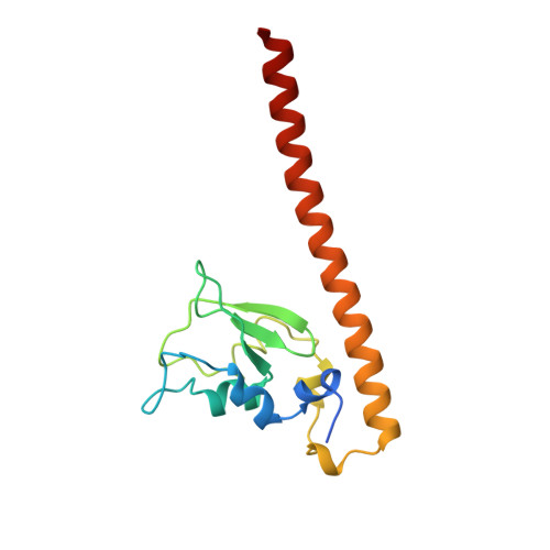
    Survivin, a subunit of the chromosome passenger complex (CPC), binds the N-terminal tail of histone H3, which is phosphorylated on T3 by Haspin kinase, and localizes the complex to the inner centromeres. We used x-ray crystallography to determine the residues of Survivin that are important in binding phosphomodified histone H3. Mutation of amino acids that interact with the histone N-terminus lowered in vitro tail binding affinity and reduced CPC recruitment to the inner centromere in cells, validating our solved structures. Phylogenetic analysis shows that nonmammalian vertebrates have two Survivin paralogues, which we name class A and B. A distinguishing feature of these paralogues is an H-to-R change in an amino acid that interacts with the histone T3 phosphate. The binding to histone tails of the human class A paralogue, which has a histidine at this position, is sensitive to changes around physiological pH, whereas Xenopus Survivin class B is less so. Our data demonstrate that Survivin paralogues have different characteristics of phosphospecific binding to threonine-3 of histone H3, providing new insight into the biology of the inner centromere.


  • Organizational Affiliation
    • Department of Biochemistry and Molecular Genetics, University of Virginia School of Medicine, Charlottesville, VA 22908, USA.

Macromolecule Content 

  • Total Structure Weight: 34.07 kDa 
  • Atom Count: 2,201 
  • Modeled Residue Count: 274 
  • Deposited Residue Count: 292 
  • Unique protein chains: 1

Macromolecules

Find similar proteins by:|  3D Structure
Entity ID: 1
MoleculeChains  Sequence LengthOrganismDetailsImage
Baculoviral IAP repeat-containing protein 5
A, B
146Homo sapiensMutation(s): 1 
Gene Names: API4BIRC5IAP4
UniProt & NIH Common Fund Data Resources
Find proteins for O15392 (Homo sapiens)
Explore O15392 
Go to UniProtKB:  O15392
PHAROS:  O15392
GTEx:  ENSG00000089685 
Entity Groups
Sequence Clusters30% Identity50% Identity70% Identity90% Identity95% Identity100% Identity
UniProt GroupO15392
Sequence Annotations
Expand
Reference Sequence

Small Molecules

Ligands 4 Unique
IDChains Name / Formula / InChI Key2D Diagram3D Interactions
PGE

Query on PGE


Download:Ideal Coordinates CCD File
F [auth A]TRIETHYLENE GLYCOL
C6 H14 O4
ZIBGPFATKBEMQZ-UHFFFAOYSA-N
PEG

Query on PEG


Download:Ideal Coordinates CCD File
E [auth A],
G [auth A]
DI(HYDROXYETHYL)ETHER
C4 H10 O3
MTHSVFCYNBDYFN-UHFFFAOYSA-N
ZN

Query on ZN


Download:Ideal Coordinates CCD File
C [auth A],
H [auth B]
ZINC ION
Zn
PTFCDOFLOPIGGS-UHFFFAOYSA-N
EDO

Query on EDO


Download:Ideal Coordinates CCD File
D [auth A]1,2-ETHANEDIOL
C2 H6 O2
LYCAIKOWRPUZTN-UHFFFAOYSA-N

Experimental Data & Validation

Experimental Data

  • Method: X-RAY DIFFRACTION
  • Resolution: 2.60 Å
  • R-Value Free:  0.224 (Depositor), 0.218 (DCC) 
  • R-Value Work:  0.189 (Depositor), 0.192 (DCC) 
  • R-Value Observed: 0.191 (Depositor) 
Space Group: C 1 2 1
Unit Cell:
Length ( Å )Angle ( ˚ )
a = 113.208α = 90
b = 71.128β = 131.43
c = 84.063γ = 90
Software Package:
Software NamePurpose
BLU-MAXdata collection
HKL-3000phasing
MOLREPphasing
REFMACrefinement
Cootmodel building
HKL-3000data reduction
HKL-3000data scaling

Structure Validation

View Full Validation Report


Entry History 

Deposition Data

Revision History  (Full details and data files)

  • Version 1.0: 2012-03-07
    Type: Initial release
  • Version 1.1: 2012-07-25
    Changes: Database references
  • Version 1.2: 2022-04-13
    Changes: Database references, Derived calculations, Structure summary
  • Version 1.3: 2023-09-13
    Changes: Data collection, Refinement description