3U2C | pdb_00003u2c

Aldose reductase in complex with NSAID-type inhibitor at 1.0 A resolution


Experimental Data Snapshot

  • Method: X-RAY DIFFRACTION
  • Resolution: 1.00 Å
  • R-Value Free: 
    0.138 (Depositor), 0.136 (DCC) 
  • R-Value Work: 
    0.113 (Depositor), 0.113 (DCC) 
  • R-Value Observed: 
    0.114 (Depositor) 

Starting Model: experimental
View more details

wwPDB Validation 3D Report Full Report

Validation slider image for 3U2C

Ligand Structure Quality Assessment 


This is version 1.2 of the entry. See complete history

Literature

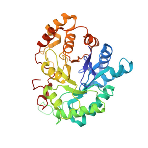
An old NSAID revisited: crystal structure of aldose reductase in complex with sulindac at 1.0 A supports a novel mechanism for its anticancer and antiproliferative effects.

Steuber, H.

(2011) ChemMedChem 6: 2155-2157

Macromolecule Content 

  • Total Structure Weight: 37.95 kDa 
  • Atom Count: 3,040 
  • Modeled Residue Count: 315 
  • Deposited Residue Count: 316 
  • Unique protein chains: 1

Macromolecules

Find similar proteins by:|  3D Structure
Entity ID: 1
MoleculeChains  Sequence LengthOrganismDetailsImage
Aldose reductase316Homo sapiensMutation(s): 0 
Gene Names: AKR1B1aldose reductaseALDR1
EC: 1.1.1.21 (PDB Primary Data), 1.1.1.372 (UniProt), 1.1.1.300 (UniProt), 1.1.1.54 (UniProt)
UniProt & NIH Common Fund Data Resources
Find proteins for P15121 (Homo sapiens)
Explore P15121 
Go to UniProtKB:  P15121
PHAROS:  P15121
GTEx:  ENSG00000085662 
Entity Groups
Sequence Clusters30% Identity50% Identity70% Identity90% Identity95% Identity100% Identity
UniProt GroupP15121
Sequence Annotations
Expand
Reference Sequence

Small Molecules

Ligands 6 Unique
IDChains Name / Formula / InChI Key2D Diagram3D Interactions
NAP

Query on NAP


Download:Ideal Coordinates CCD File
B [auth A]NADP NICOTINAMIDE-ADENINE-DINUCLEOTIDE PHOSPHATE
C21 H28 N7 O17 P3
XJLXINKUBYWONI-NNYOXOHSSA-N
SUZ

Query on SUZ


Download:Ideal Coordinates CCD File
G [auth A][(1Z)-5-fluoro-2-methyl-1-{4-[methylsulfinyl]benzylidene}-1H-inden-3-yl]acetic acid
C20 H17 F O3 S
MLKXDPUZXIRXEP-LQVWSKNFSA-N
PG4

Query on PG4


Download:Ideal Coordinates CCD File
I [auth A]TETRAETHYLENE GLYCOL
C8 H18 O5
UWHCKJMYHZGTIT-UHFFFAOYSA-N
CIT

Query on CIT


Download:Ideal Coordinates CCD File
F [auth A]CITRIC ACID
C6 H8 O7
KRKNYBCHXYNGOX-UHFFFAOYSA-N
PG5

Query on PG5


Download:Ideal Coordinates CCD File
D [auth A],
E [auth A]
1-METHOXY-2-[2-(2-METHOXY-ETHOXY]-ETHANE
C8 H18 O4
YFNKIDBQEZZDLK-UHFFFAOYSA-N
PEG

Query on PEG


Download:Ideal Coordinates CCD File
C [auth A],
H [auth A]
DI(HYDROXYETHYL)ETHER
C4 H10 O3
MTHSVFCYNBDYFN-UHFFFAOYSA-N
Binding Affinity Annotations 
IDSourceBinding Affinity
SUZ BindingDB:  3U2C IC50: 293 (nM) from 1 assay(s)

Experimental Data & Validation

Experimental Data

  • Method: X-RAY DIFFRACTION
  • Resolution: 1.00 Å
  • R-Value Free:  0.138 (Depositor), 0.136 (DCC) 
  • R-Value Work:  0.113 (Depositor), 0.113 (DCC) 
  • R-Value Observed: 0.114 (Depositor) 
Space Group: P 1 21 1
Unit Cell:
Length ( Å )Angle ( ˚ )
a = 47.095α = 90
b = 67.207β = 92.38
c = 49.419γ = 90
Software Package:
Software NamePurpose
MOLREPphasing
SHELXL-97refinement
XDSdata reduction
XDSdata scaling

Structure Validation

View Full Validation Report


Ligand Structure Quality Assessment 


Entry History 

Deposition Data

  • Released Date: 2011-11-02 
  • Deposition Author(s): Steuber, H.

Revision History  (Full details and data files)

  • Version 1.0: 2011-11-02
    Type: Initial release
  • Version 1.1: 2013-07-03
    Changes: Database references
  • Version 1.2: 2023-09-13
    Changes: Data collection, Database references, Derived calculations, Refinement description