3TYY

Crystal Structure of Human Lamin-B1 Coil 2 Segment


Experimental Data Snapshot

  • Method: X-RAY DIFFRACTION
  • Resolution: 2.40 Å
  • R-Value Free: 0.261 
  • R-Value Work: 0.235 
  • R-Value Observed: 0.236 

wwPDB Validation   3D Report Full Report


This is version 1.3 of the entry. See complete history


Literature

Crystal structures of the coil 2B fragment and the globular tail domain of human lamin B1.

Ruan, J.Xu, C.Bian, C.Lam, R.Wang, J.P.Kania, J.Min, J.Zang, J.

(2012) FEBS Lett 586: 314-318

  • DOI: https://doi.org/10.1016/j.febslet.2012.01.007
  • Primary Citation of Related Structures:  
    3TYY, 3UMN

  • PubMed Abstract: 

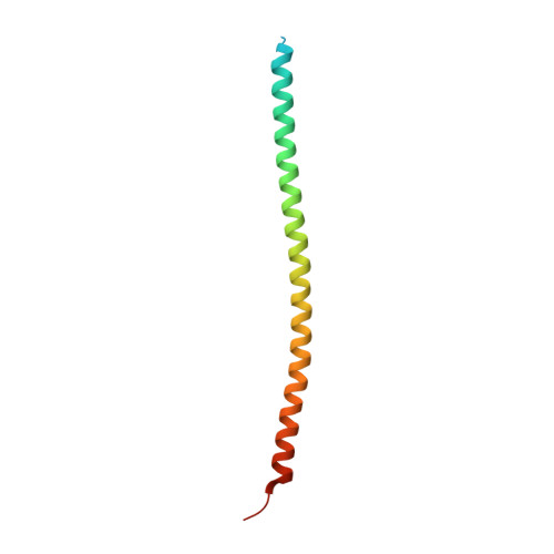
    We present here the crystal structures of human lamin B1 globular tail domain and coiled 2B domain, which adopt similar folds to Ig-like domain and coiled-coil domain of lamin A, respectively. Despite the overall similarity, we found an extra intermolecular disulfide bond in the lamin B1 coil 2B domain, which does not exist in lamin A/C. In addition, the structural analysis indicates that interactions at the lamin B1 homodimer interface are quite different from those of lamin A/C. Thus our research not only reveals the diversely formed homodimers among lamin family members, but also sheds light on understanding the important roles of lamin B1 in forming the nuclear lamina matrix.


  • Organizational Affiliation

    Hefei National Laboratory for Physical Sciences at Microscale and School of Life Sciences, University of Science and Technology of China, Hefei, Anhui, People's Republic of China.


Macromolecules
Find similar proteins by:  (by identity cutoff)  |  3D Structure
Entity ID: 1
MoleculeChains Sequence LengthOrganismDetailsImage
Lamin-B1
A, B
95Homo sapiensMutation(s): 0 
Gene Names: LMN2LMNBLMNB1
UniProt & NIH Common Fund Data Resources
Find proteins for P20700 (Homo sapiens)
Explore P20700 
Go to UniProtKB:  P20700
PHAROS:  P20700
GTEx:  ENSG00000113368 
Entity Groups  
Sequence Clusters30% Identity50% Identity70% Identity90% Identity95% Identity100% Identity
UniProt GroupP20700
Sequence Annotations
Expand
  • Reference Sequence
Experimental Data & Validation

Experimental Data

  • Method: X-RAY DIFFRACTION
  • Resolution: 2.40 Å
  • R-Value Free: 0.261 
  • R-Value Work: 0.235 
  • R-Value Observed: 0.236 
  • Space Group: P 32 2 1
Unit Cell:
Length ( Å )Angle ( ˚ )
a = 45.982α = 90
b = 45.982β = 90
c = 203.231γ = 120
Software Package:
Software NamePurpose
DENZOdata reduction
SCALEPACKdata scaling
PHASERphasing
REFMACrefinement
PDB_EXTRACTdata extraction
Locallydata collection
HKL-2000data reduction
HKL-2000data scaling

Structure Validation

View Full Validation Report


Entry History 

Deposition Data

Revision History  (Full details and data files)

  • Version 1.0: 2011-10-05
    Type: Initial release
  • Version 1.1: 2012-03-07
    Changes: Database references
  • Version 1.2: 2017-11-08
    Changes: Refinement description
  • Version 1.3: 2023-09-13
    Changes: Data collection, Database references, Refinement description