3TSZ

crystal structure of PDZ3-SH3-GUK core module from human ZO-1 in complex with 12mer peptide from human JAM-A cytoplasmic tail


Experimental Data Snapshot

  • Method: X-RAY DIFFRACTION
  • Resolution: 2.50 Å
  • R-Value Free: 0.309 
  • R-Value Work: 0.240 
  • R-Value Observed: 0.247 

wwPDB Validation   3D Report Full Report


This is version 1.3 of the entry. See complete history


Literature

The Src Homology 3 Domain Is Required for Junctional Adhesion Molecule Binding to the Third PDZ Domain of the Scaffolding Protein ZO-1.

Nomme, J.Fanning, A.S.Caffrey, M.Lye, M.F.Anderson, J.M.Lavie, A.

(2011) J Biol Chem 286: 43352-43360

  • DOI: https://doi.org/10.1074/jbc.M111.304089
  • Primary Citation of Related Structures:  
    3TSV, 3TSW, 3TSZ

  • PubMed Abstract: 

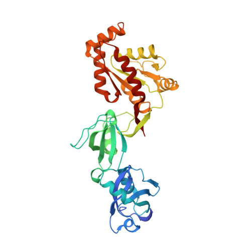
    Tight junctions are cell-cell contacts that regulate the paracellular flux of solutes and prevent pathogen entry across cell layers. The assembly and permeability of this barrier are dependent on the zonula occludens (ZO) membrane-associated guanylate kinase (MAGUK) proteins ZO-1, -2, and -3. MAGUK proteins are characterized by a core motif of protein-binding domains that include a PDZ domain, a Src homology 3 (SH3) domain, and a region of homology to guanylate kinase (GUK); the structure of this core motif has never been determined for any MAGUK. To better understand how ZO proteins organize the assembly of protein complexes we have crystallized the entire PDZ3-SH3-GUK core motif of ZO-1. We have also crystallized this core motif in complex with the cytoplasmic tail of the ZO-1 PDZ3 ligand, junctional adhesion molecule A (JAM-A) to determine how the activity of different domains is coordinated. Our study shows a new feature for PDZ class II ligand binding that implicates the two highly conserved Phe(-2) and Ser(-3) residues of JAM. Our x-ray structures and NMR experiments also show for the first time a role for adjacent domains in the binding of ligands to PDZ domains in the MAGUK proteins family.


  • Organizational Affiliation

    Department of Biochemistry and Molecular Genetics, University of Illinois, Chicago, Illinois 60607, USA.


Macromolecules
Find similar proteins by:  (by identity cutoff)  |  3D Structure
Entity ID: 1
MoleculeChains Sequence LengthOrganismDetailsImage
Tight junction protein ZO-1391Homo sapiensMutation(s): 0 
Gene Names: TJP1ZO1
UniProt & NIH Common Fund Data Resources
Find proteins for Q07157 (Homo sapiens)
Explore Q07157 
Go to UniProtKB:  Q07157
PHAROS:  Q07157
GTEx:  ENSG00000104067 
Entity Groups  
Sequence Clusters30% Identity50% Identity70% Identity90% Identity95% Identity100% Identity
UniProt GroupQ07157
Sequence Annotations
Expand
  • Reference Sequence

Find similar proteins by:  Sequence   |   3D Structure  

Entity ID: 2
MoleculeChains Sequence LengthOrganismDetailsImage
Junctional adhesion molecule A12Homo sapiensMutation(s): 0 
UniProt & NIH Common Fund Data Resources
Find proteins for Q9Y624 (Homo sapiens)
Explore Q9Y624 
Go to UniProtKB:  Q9Y624
PHAROS:  Q9Y624
GTEx:  ENSG00000158769 
Entity Groups  
Sequence Clusters30% Identity50% Identity70% Identity90% Identity95% Identity100% Identity
UniProt GroupQ9Y624
Sequence Annotations
Expand
  • Reference Sequence
Experimental Data & Validation

Experimental Data

  • Method: X-RAY DIFFRACTION
  • Resolution: 2.50 Å
  • R-Value Free: 0.309 
  • R-Value Work: 0.240 
  • R-Value Observed: 0.247 
  • Space Group: P 1 21 1
Unit Cell:
Length ( Å )Angle ( ˚ )
a = 50.5α = 90
b = 49.7β = 101.1
c = 91.8γ = 90
Software Package:
Software NamePurpose
SERGUIdata collection
PHASERphasing
REFMACrefinement
XDSdata reduction
XDSdata scaling

Structure Validation

View Full Validation Report


Entry History 

Deposition Data

Revision History  (Full details and data files)

  • Version 1.0: 2011-10-12
    Type: Initial release
  • Version 1.1: 2011-11-16
    Changes: Database references
  • Version 1.2: 2011-12-28
    Changes: Database references
  • Version 1.3: 2023-09-13
    Changes: Data collection, Database references, Refinement description