Control of Substrate Specificity by a Single Active Site Residue of the KsgA Methyltransferase.
O'Farrell, H.C., Musayev, F.N., Scarsdale, J.N., Rife, J.P.(2012) Biochemistry 51: 466-474
- PubMed: 22142337 Search on PubMed
- DOI: https://doi.org/10.1021/bi201539j
- Primary Citation Related Structures:
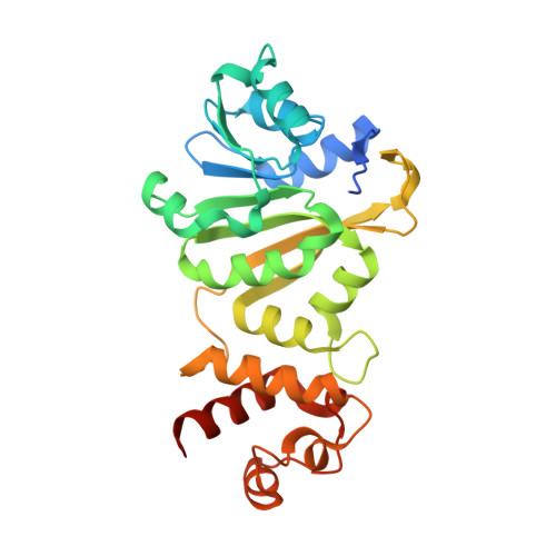
3TPZ - PubMed Abstract:
The KsgA methyltransferase is universally conserved and plays a key role in regulating ribosome biogenesis. KsgA has a complex reaction mechanism, transferring a total of four methyl groups onto two separate adenosine residues, A1518 and A1519, in the small subunit rRNA. This means that the active site pocket must accept both adenosine and N(6)-methyladenosine as substrates to catalyze formation of the final product N(6),N(6)-dimethyladenosine. KsgA is related to DNA adenosine methyltransferases, which transfer only a single methyl group to their target adenosine residue. We demonstrate that part of the discrimination between mono- and dimethyltransferase activity lies in a single residue in the active site, L114; this residue is part of a conserved motif, known as motif IV, which is common to a large group of S-adenosyl-L-methionine-dependent methyltransferases. Mutation of the leucine to a proline mimics the sequence found in DNA methyltransferases. The L114P mutant of KsgA shows diminished overall activity, and its ability to methylate the N(6)-methyladenosine intermediate to produce N(6),N(6)-dimethyladenosine is impaired; this is in contrast to a second active site mutation, N113A, which diminishes activity to a level comparable to L114P without affecting the methylation of N(6)-methyladenosine. We discuss the implications of this work for understanding the mechanism of KsgA's multiple catalytic steps.
- Department of Physiology and Molecular Biophysics, Virginia Commonwealth University, Richmond, Virginia 23219, United States.
Organizational Affiliation: