3TJ8 | pdb_00003tj8

Crystal structure of Helicobacter pylori UreE bound to Ni2+


Experimental Data Snapshot

  • Method: X-RAY DIFFRACTION
  • Resolution: 1.59 Å
  • R-Value Free: 
    0.211 (Depositor), 0.209 (DCC) 
  • R-Value Work: 
    0.185 (Depositor), 0.184 (DCC) 
  • R-Value Observed: 
    0.186 (Depositor) 

Starting Model: experimental
View more details

wwPDB Validation 3D Report Full Report

Validation slider image for 3TJ8

This is version 1.3 of the entry. See complete history

Literature

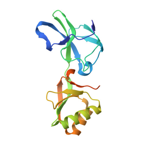
Crystallographic and X-ray absorption spectroscopic characterization of Helicobacter pylori UreE bound to Ni2+ and Zn2+ reveals a role for the disordered C-terminal arm in metal trafficking.

Banaszak, K.Martin-Diaconescu, V.Bellucci, M.Zambelli, B.Rypniewski, W.Maroney, M.J.Ciurli, S.

(2012) Biochem J 441: 1017-1026

  • DOI: https://doi.org/10.1042/BJ20111659
  • Primary Citation Related Structures: 
    3TJ8, 3TJ9, 3TJA

  • PubMed Abstract: 

    The survival and growth of the pathogen Helicobacter pylori in the gastric acidic environment is ensured by the activity of urease, an enzyme containing two essential Ni²⁺ ions in the active site. The metallo-chaperone UreE facilitates in vivo Ni²⁺ insertion into the apoenzyme. Crystals of apo-HpUreE (H. pylori UreE) and its Ni⁺- and Zn⁺-bound forms were obtained from protein solutions in the absence and presence of the metal ions. The crystal structures of the homodimeric protein, determined at 2.00 Å (apo), 1.59 Å (Ni²⁺) and 2.52 Å (Zn²⁺) resolution, show the conserved proximal and solvent-exposed His¹⁰² residues from two adjacent monomers invariably involved in metal binding. The C-terminal regions of the apoprotein are disordered in the crystal, but acquire significant ordering in the presence of the metal ions due to the binding of His¹⁵². The analysis of X-ray absorption spectral data obtained using solutions of Ni²⁺- and Zn²⁺-bound HpUreE provided accurate information of the metal-ion environment in the absence of solid-state effects. These results reveal the role of the histidine residues at the protein C-terminus in metal-ion binding, and the mutual influence of protein framework and metal-ion stereo-electronic properties in establishing co-ordination number and geometry leading to metal selectivity.


  • Organizational Affiliation
    • Institute of Bioorganic Chemistry, Polish Academy of Sciences, Noskowskiego 12/14, 61-704 Poznan, Poland.

Macromolecule Content 

  • Total Structure Weight: 39.39 kDa 
  • Atom Count: 2,587 
  • Modeled Residue Count: 301 
  • Deposited Residue Count: 340 
  • Unique protein chains: 1

Macromolecules

Find similar proteins by:|  3D Structure
Entity ID: 1
MoleculeChains  Sequence LengthOrganismDetailsImage
Urease accessory protein ureE
A, B
170Helicobacter pyloriMutation(s): 0 
Gene Names: ureEHP_0070
UniProt
Find proteins for Q09064 (Helicobacter pylori (strain ATCC 700392 / 26695))
Explore Q09064 
Go to UniProtKB:  Q09064
Entity Groups
Sequence Clusters30% Identity50% Identity70% Identity90% Identity95% Identity100% Identity
UniProt GroupQ09064
Sequence Annotations
Expand
Reference Sequence

Small Molecules

Ligands 2 Unique
IDChains Name / Formula / InChI Key2D Diagram3D Interactions
NI

Query on NI


Download:Ideal Coordinates CCD File
G [auth B]NICKEL (II) ION
Ni
VEQPNABPJHWNSG-UHFFFAOYSA-N
FMT

Query on FMT


Download:Ideal Coordinates CCD File
C [auth A]
D [auth A]
E [auth A]
F [auth A]
H [auth B]
C [auth A],
D [auth A],
E [auth A],
F [auth A],
H [auth B],
I [auth B],
J [auth B],
K [auth B],
L [auth B],
M [auth B]
FORMIC ACID
C H2 O2
BDAGIHXWWSANSR-UHFFFAOYSA-N

Experimental Data & Validation

Experimental Data

  • Method: X-RAY DIFFRACTION
  • Resolution: 1.59 Å
  • R-Value Free:  0.211 (Depositor), 0.209 (DCC) 
  • R-Value Work:  0.185 (Depositor), 0.184 (DCC) 
  • R-Value Observed: 0.186 (Depositor) 
Space Group: C 2 2 21
Unit Cell:
Length ( Å )Angle ( ˚ )
a = 67.552α = 90
b = 117.145β = 90
c = 98.665γ = 90
Software Package:
Software NamePurpose
MxCuBEdata collection
PHASERphasing
REFMACrefinement
HKL-2000data reduction
HKL-2000data scaling

Structure Validation

View Full Validation Report


Entry History 

Deposition Data

Revision History  (Full details and data files)

  • Version 1.0: 2011-11-02
    Type: Initial release
  • Version 1.1: 2012-02-01
    Changes: Database references
  • Version 1.2: 2024-02-28
    Changes: Data collection, Database references, Derived calculations
  • Version 1.3: 2024-04-03
    Changes: Refinement description