


Structure of subunit A mutants H156A, N157A and N157T of the A1AO ATP synthase
Tadwal, V.S., Manimekalai, M.S.S., Gruber, G.To be published.
Experimental Data Snapshot
Starting Model: experimental
View more details
Entity ID: 1 | |||||
|---|---|---|---|---|---|
| Molecule | Chains | Sequence Length | Organism | Details | Image |
| V-type ATP synthase beta chain | 460 | Methanosarcina mazei Go1 | Mutation(s): 1 Gene Names: ahaB, atpB, MM_0779 EC: 3.6.3.14 | 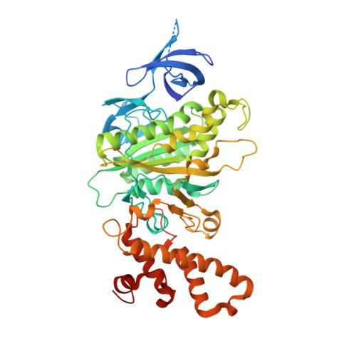 | |
UniProt | |||||
Entity Groups | |||||
| Sequence Clusters | 30% Identity50% Identity70% Identity90% Identity95% Identity100% Identity | ||||
| UniProt Group | Q60187 | ||||
Sequence AnnotationsExpand | |||||
Reference Sequence | |||||
| Ligands 6 Unique | |||||
|---|---|---|---|---|---|
| ID | Chains | Name / Formula / InChI Key | 2D Diagram | 3D Interactions | |
| 1PE Download:Ideal Coordinates CCD File | P [auth B] | PENTAETHYLENE GLYCOL C10 H22 O6 JLFNLZLINWHATN-UHFFFAOYSA-N |  | ||
| AES Download:Ideal Coordinates CCD File | O [auth B] | 4-(2-AMINOETHYL)BENZENESULFONYL FLUORIDE C8 H10 F N O2 S MGSKVZWGBWPBTF-UHFFFAOYSA-N |  | ||
| PG4 Download:Ideal Coordinates CCD File | N [auth B] | TETRAETHYLENE GLYCOL C8 H18 O5 UWHCKJMYHZGTIT-UHFFFAOYSA-N |  | ||
| PG0 Download:Ideal Coordinates CCD File | I [auth A] | 2-(2-METHOXYETHOXY)ETHANOL C5 H12 O3 SBASXUCJHJRPEV-UHFFFAOYSA-N |  | ||
| GOL Download:Ideal Coordinates CCD File | C [auth A] D [auth A] E [auth A] F [auth A] G [auth A] | GLYCEROL C3 H8 O3 PEDCQBHIVMGVHV-UHFFFAOYSA-N |  | ||
| CL Download:Ideal Coordinates CCD File | R [auth B] | CHLORIDE ION Cl VEXZGXHMUGYJMC-UHFFFAOYSA-M |  | ||
| Length ( Å ) | Angle ( ˚ ) |
|---|---|
| a = 73.715 | α = 90 |
| b = 95.77 | β = 90 |
| c = 130.158 | γ = 90 |
| Software Name | Purpose |
|---|---|
| PHASER | phasing |
| REFMAC | refinement |
| PDB_EXTRACT | data extraction |
| HKL-2000 | data collection |
| HKL-2000 | data reduction |
| HKL-2000 | data scaling |


