3TBJ

The 1.7A crystal structure of Actibind a T2 ribonucleases as antitumorigenic agents


Experimental Data Snapshot

  • Method: X-RAY DIFFRACTION
  • Resolution: 1.80 Å
  • R-Value Free: 0.187 
  • R-Value Work: 0.154 
  • R-Value Observed: 0.155 

wwPDB Validation   3D Report Full Report


Ligand Structure Quality Assessment 


This is version 2.0 of the entry. See complete history


Literature

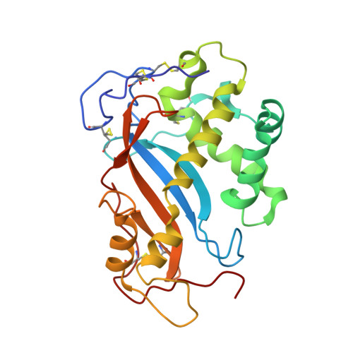
The 1.8 A crystal structure of ACTIBIND suggests a mode of action for T2 ribonucleases as antitumorigenic agents.

de Leeuw, M.Gonzalez, A.Lanir, A.Roiz, L.Smirnoff, P.Schwartz, B.Shoseyov, O.Almog, O.

(2012) J Med Chem 55: 1013-1020

  • DOI: https://doi.org/10.1021/jm1015507
  • Primary Citation of Related Structures:  
    3TBJ

  • PubMed Abstract: 

    ACTIBIND and its human homologue RNASET2 are T2 ribonucleases (RNases). RNases are ubiquitous and efficient enzymes that hydrolyze RNA to 3' mononucleotides and also possess antitumorigenic and antiangiogenic activities. Previously, we have shown that ACTIBIND and RNASET2 bind actin and interfere with the cytoskeletal network structure, thereby inhibiting cell motility and invasiveness in cancer and in endothelial cells. We also showed that ACTIBIND binds actin in a molar ratio of 1:2. Here, we further characterize ACTIBIND and determine its crystal structure at 1.8 Å resolution, which enables us to propose two structural elements that create binding sites to actin. We suggest that each of these binding sites is composed of one cysteine residue and one conserved amino acid region. These binding sites possibly interfere with the cytoskeleton network structure and as such may be responsible for the antitumorigenic and antiangiogenic activities of ACTIBIND and its human analogue RNASET2.


  • Organizational Affiliation

    Department of Clinical Biochemistry, Faculty of Health Sciences, Ben-Gurion University of the Negev, Beer Sheva 84105, Israel.


Macromolecules
Find similar proteins by:  (by identity cutoff)  |  3D Structure
Entity ID: 1
MoleculeChains Sequence LengthOrganismDetailsImage
Actibind237Aspergillus nigerMutation(s): 0 
UniProt
Find proteins for Q45U61 (Aspergillus niger)
Explore Q45U61 
Go to UniProtKB:  Q45U61
Entity Groups  
Sequence Clusters30% Identity50% Identity70% Identity90% Identity95% Identity100% Identity
UniProt GroupQ45U61
Sequence Annotations
Expand
  • Reference Sequence
Oligosaccharides

Help

Entity ID: 2
MoleculeChains Length2D Diagram Glycosylation3D Interactions
2-acetamido-2-deoxy-beta-D-glucopyranose-(1-4)-2-acetamido-2-deoxy-beta-D-glucopyranose
B
2N-Glycosylation
Glycosylation Resources
GlyTouCan:  G42666HT
GlyCosmos:  G42666HT
GlyGen:  G42666HT
Experimental Data & Validation

Experimental Data

  • Method: X-RAY DIFFRACTION
  • Resolution: 1.80 Å
  • R-Value Free: 0.187 
  • R-Value Work: 0.154 
  • R-Value Observed: 0.155 
  • Space Group: P 32 2 1
Unit Cell:
Length ( Å )Angle ( ˚ )
a = 78.457α = 90
b = 78.457β = 90
c = 103.708γ = 120
Software Package:
Software NamePurpose
SCALAdata scaling
MOLREPphasing
REFMACrefinement
PDB_EXTRACTdata extraction
Blu-Icedata collection

Structure Validation

View Full Validation Report


Ligand Structure Quality Assessment 


Entry History 

Deposition Data

Revision History  (Full details and data files)

  • Version 1.0: 2012-08-22
    Type: Initial release
  • Version 2.0: 2020-07-29
    Type: Remediation
    Reason: Carbohydrate remediation
    Changes: Atomic model, Data collection, Derived calculations, Structure summary