3TAX | pdb_00003tax

A Neutral Diphosphate Mimic Crosslinks the Active Site of Human O-GlcNAc Transferase


Experimental Data Snapshot

  • Method: X-RAY DIFFRACTION
  • Resolution: 1.88 Å
  • R-Value Free: 
    0.237 (Depositor), 0.237 (DCC) 
  • R-Value Work: 
    0.219 (Depositor), 0.217 (DCC) 
  • R-Value Observed: 
    0.220 (Depositor) 

Starting Model: experimental
View more details

wwPDB Validation 3D Report Full Report

Validation slider image for 3TAX

Ligand Structure Quality Assessment 


This is version 1.3 of the entry. See complete history

Literature

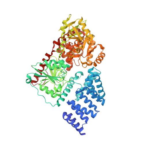
A neutral diphosphate mimic crosslinks the active site of human O-GlcNAc transferase.

Jiang, J.Lazarus, M.B.Pasquina, L.Sliz, P.Walker, S.

(2011) Nat Chem Biol 8: 72-77

  • DOI: https://doi.org/10.1038/nchembio.711
  • Primary Citation Related Structures: 
    3TAX

  • PubMed Abstract: 

    Glycosyltransferases (Gtfs) catalyze the formation of a diverse array of glycoconjugates. Small-molecule inhibitors to manipulate Gtf activity in cells have long been sought as tools for understanding Gtf function. Success has been limited because of challenges in designing inhibitors that mimic the negatively charged diphosphate substrates. Here we report the mechanism of action of a small molecule that inhibits O-linked N-acetylglucosamine transferase (OGT), an essential human enzyme that modulates cell signaling pathways by catalyzing a unique intracellular post-translational modification, β-O-GlcNAcylation. The molecule contains a five-heteroatom dicarbamate core that functions as a neutral diphosphate mimic. One dicarbamate carbonyl reacts with an essential active site lysine that anchors the diphosphate of the nucleotide-sugar substrate. A nearby cysteine then reacts with the lysine adduct to form a carbonyl crosslink in the OGT active site. Though this unprecedented double-displacement mechanism reflects the unique architecture of the OGT active site, related dicarbamate scaffolds may inhibit other enzymes that bind nucleotide-containing substrates.


  • Organizational Affiliation
    • Department of Microbiology and Immunobiology, Harvard Medical School, Boston, Massachusetts, USA.

Macromolecule Content 

  • Total Structure Weight: 166 kDa 
  • Atom Count: 12,326 
  • Modeled Residue Count: 1,418 
  • Deposited Residue Count: 1,474 
  • Unique protein chains: 2

Macromolecules

Find similar proteins by:|  3D Structure
Entity ID: 1
MoleculeChains  Sequence LengthOrganismDetailsImage
UDP-N-acetylglucosamine--peptide N-acetylglucosaminyltransferase 110 kDa subunit
A, C
723Homo sapiensMutation(s): 0 
Gene Names: OGT
EC: 2.4.1 (PDB Primary Data), 2.4.1.255 (UniProt)
UniProt & NIH Common Fund Data Resources
Find proteins for O15294 (Homo sapiens)
Explore O15294 
Go to UniProtKB:  O15294
PHAROS:  O15294
GTEx:  ENSG00000147162 
Entity Groups
Sequence Clusters30% Identity50% Identity70% Identity90% Identity95% Identity100% Identity
UniProt GroupO15294
Sequence Annotations
Expand
Reference Sequence
Find similar proteins by:  Sequence   |   3D Structure  
Entity ID: 2
MoleculeChains  Sequence LengthOrganismDetailsImage
Casein kinase II subunit alpha
B, D
14Homo sapiensMutation(s): 0 
EC: 2.7.11.1
UniProt & NIH Common Fund Data Resources
Find proteins for P68400 (Homo sapiens)
Explore P68400 
Go to UniProtKB:  P68400
PHAROS:  P68400
GTEx:  ENSG00000101266 
Entity Groups
Sequence Clusters30% Identity50% Identity70% Identity90% Identity95% Identity100% Identity
UniProt GroupP68400
Sequence Annotations
Expand
Reference Sequence

Small Molecules

Ligands 3 Unique
IDChains Name / Formula / InChI Key2D Diagram3D Interactions
UDP

Query on UDP


Download:Ideal Coordinates CCD File
E [auth A],
I [auth C]
URIDINE-5'-DIPHOSPHATE
C9 H14 N2 O12 P2
XCCTYIAWTASOJW-XVFCMESISA-N
SO4

Query on SO4


Download:Ideal Coordinates CCD File
G [auth A],
H [auth B],
J [auth C],
L [auth D]
SULFATE ION
O4 S
QAOWNCQODCNURD-UHFFFAOYSA-L
FOR

Query on FOR


Download:Ideal Coordinates CCD File
F [auth A],
K [auth C]
FORMYL GROUP
C H2 O
WSFSSNUMVMOOMR-UHFFFAOYSA-N

Experimental Data & Validation

Experimental Data

  • Method: X-RAY DIFFRACTION
  • Resolution: 1.88 Å
  • R-Value Free:  0.237 (Depositor), 0.237 (DCC) 
  • R-Value Work:  0.219 (Depositor), 0.217 (DCC) 
  • R-Value Observed: 0.220 (Depositor) 
Space Group: I 1 2 1
Unit Cell:
Length ( Å )Angle ( ˚ )
a = 98.6α = 90
b = 136.64β = 103.02
c = 153.319γ = 90
Software Package:
Software NamePurpose
ADSCdata collection
PHASERphasing
BUSTERrefinement
iMOSFLMdata reduction
SCALAdata scaling

Structure Validation

View Full Validation Report


Ligand Structure Quality Assessment 


Entry History 

Deposition Data

Revision History  (Full details and data files)

  • Version 1.0: 2011-11-16
    Type: Initial release
  • Version 1.1: 2011-12-28
    Changes: Database references
  • Version 1.2: 2023-09-13
    Changes: Data collection, Database references, Derived calculations, Refinement description
  • Version 1.3: 2024-10-30
    Changes: Structure summary