3SYN | pdb_00003syn

Crystal structure of FlhF in complex with its activator


Experimental Data Snapshot

  • Method: X-RAY DIFFRACTION
  • Resolution: 3.06 Å
  • R-Value Free: 
    0.239 (Depositor), 0.229 (DCC) 
  • R-Value Work: 
    0.181 (Depositor), 0.174 (DCC) 
  • R-Value Observed: 
    0.184 (Depositor) 

Starting Model: experimental
View more details

wwPDB Validation 3D Report Full Report

Validation slider image for 3SYN

Ligand Structure Quality Assessment 


This is version 1.2 of the entry. See complete history

Literature

Structural basis for the molecular evolution of SRP-GTPase activation by protein.

Bange, G.Kummerer, N.Grudnik, P.Lindner, R.Petzold, G.Kressler, D.Hurt, E.Wild, K.Sinning, I.

(2011) Nat Struct Mol Biol 18: 1376-1380

  • DOI: https://doi.org/10.1038/nsmb.2141
  • Primary Citation Related Structures: 
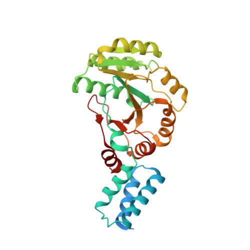
    3SYN

  • PubMed Abstract: 

    Small G proteins have key roles in signal transduction pathways. They are switched from the signaling 'on' to the non-signaling 'off' state when GTPase-activating proteins (GAPs) provide a catalytic residue. The ancient signal recognition particle (SRP)-type GTPases form GTP-dependent homo- and heterodimers and deviate from the canonical switch paradigm in that no GAPs have been identified. Here we show that the YlxH protein activates the SRP-GTPase FlhF. The crystal structure of the Bacillus subtilis FlhF-effector complex revealed that the effector does not contribute a catalytic residue but positions the catalytic machinery already present in SRP-GTPases. We provide a general concept that might also apply to the RNA-driven activation of the universally conserved, co-translational protein-targeting machinery comprising the SRP-GTPases Ffh and FtsY. Our study exemplifies the evolutionary transition from RNA- to protein-driven activation in SRP-GTPases and suggests that the current view on SRP-mediated protein targeting is incomplete.


  • Organizational Affiliation
    • Heidelberg University Biochemistry Center, Heidelberg, Germany. gert.bange@bzh.uni-heidelberg.de

Macromolecule Content 

  • Total Structure Weight: 148.76 kDa 
  • Atom Count: 9,010 
  • Modeled Residue Count: 1,096 
  • Deposited Residue Count: 1,276 
  • Unique protein chains: 2

Macromolecules

Find similar proteins by:|  3D Structure
Entity ID: 1
MoleculeChains  Sequence LengthOrganismDetailsImage
Flagellar biosynthesis protein flhF
A, B, C, D
296Bacillus subtilisMutation(s): 0 
Gene Names: flhFBSU16400
UniProt
Find proteins for Q01960 (Bacillus subtilis (strain 168))
Explore Q01960 
Go to UniProtKB:  Q01960
Entity Groups
Sequence Clusters30% Identity50% Identity70% Identity90% Identity95% Identity100% Identity
UniProt GroupQ01960
Sequence Annotations
Expand
Reference Sequence
Find similar proteins by:  Sequence   |   3D Structure  
Entity ID: 2
MoleculeChains  Sequence LengthOrganismDetailsImage
ATP-binding protein YlxH
E, F, G, H
23Bacillus subtilisMutation(s): 0 
UniProt
Find proteins for P40742 (Bacillus subtilis (strain 168))
Explore P40742 
Go to UniProtKB:  P40742
Entity Groups
Sequence Clusters30% Identity50% Identity70% Identity90% Identity95% Identity100% Identity
UniProt GroupP40742
Sequence Annotations
Expand
Reference Sequence

Small Molecules

Ligands 4 Unique
IDChains Name / Formula / InChI Key2D Diagram3D Interactions
GDP

Query on GDP


Download:Ideal Coordinates CCD File
DA [auth D],
I [auth A],
Q [auth B],
W [auth C]
GUANOSINE-5'-DIPHOSPHATE
C10 H15 N5 O11 P2
QGWNDRXFNXRZMB-UUOKFMHZSA-N
SO4

Query on SO4


Download:Ideal Coordinates CCD File
AA [auth C]
BA [auth C]
CA [auth C]
GA [auth D]
HA [auth D]
AA [auth C],
BA [auth C],
CA [auth C],
GA [auth D],
HA [auth D],
IA [auth D],
L [auth A],
M [auth A],
N [auth A],
O [auth A],
P [auth A],
T [auth B],
U [auth B],
V [auth B],
Z [auth C]
SULFATE ION
O4 S
QAOWNCQODCNURD-UHFFFAOYSA-L
AF3

Query on AF3


Download:Ideal Coordinates CCD File
FA [auth D],
K [auth A],
S [auth B],
Y [auth C]
ALUMINUM FLUORIDE
Al F3
KLZUFWVZNOTSEM-UHFFFAOYSA-K
MG

Query on MG


Download:Ideal Coordinates CCD File
EA [auth D],
J [auth A],
R [auth B],
X [auth C]
MAGNESIUM ION
Mg
JLVVSXFLKOJNIY-UHFFFAOYSA-N

Experimental Data & Validation

Experimental Data

  • Method: X-RAY DIFFRACTION
  • Resolution: 3.06 Å
  • R-Value Free:  0.239 (Depositor), 0.229 (DCC) 
  • R-Value Work:  0.181 (Depositor), 0.174 (DCC) 
  • R-Value Observed: 0.184 (Depositor) 
Space Group: P 1
Unit Cell:
Length ( Å )Angle ( ˚ )
a = 50.79α = 92.26
b = 63.36β = 100.96
c = 114.5γ = 94.25
Software Package:
Software NamePurpose
ADSCdata collection
PHENIXmodel building
PHENIXrefinement
MOSFLMdata reduction
SCALAdata scaling
PHENIXphasing

Structure Validation

View Full Validation Report


Ligand Structure Quality Assessment 


Entry History 

Deposition Data

Revision History  (Full details and data files)

  • Version 1.0: 2011-11-09
    Type: Initial release
  • Version 1.1: 2012-05-23
    Changes: Database references
  • Version 1.2: 2023-09-13
    Changes: Data collection, Database references, Derived calculations, Refinement description