3SLJ | pdb_00003slj

Pre-cleavage Structure of the Autotransporter EspP - N1023A mutant


Experimental Data Snapshot

  • Method: X-RAY DIFFRACTION
  • Resolution: 2.48 Å
  • R-Value Free: 
    0.237 (Depositor), 0.230 (DCC) 
  • R-Value Work: 
    0.199 (Depositor), 0.191 (DCC) 
  • R-Value Observed: 
    0.201 (Depositor) 

Starting Model: experimental
View more details

wwPDB Validation 3D Report Full Report

Validation slider image for 3SLJ

Ligand Structure Quality Assessment 


This is version 1.4 of the entry. See complete history

Literature

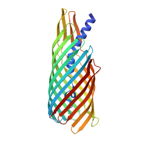
Molecular basis for the activation of a catalytic asparagine residue in a self-cleaving bacterial autotransporter.

Barnard, T.J.Gumbart, J.Peterson, J.H.Noinaj, N.Easley, N.C.Dautin, N.Kuszak, A.J.Tajkhorshid, E.Bernstein, H.D.Buchanan, S.K.

(2012) J Mol Biology 415: 128-142

  • DOI: https://doi.org/10.1016/j.jmb.2011.10.049
  • Primary Citation Related Structures: 
    3SLJ, 3SLO, 3SLT

  • PubMed Abstract: 

    Autotransporters are secreted proteins produced by pathogenic Gram-negative bacteria. They consist of a membrane-embedded β-domain and an extracellular passenger domain that is sometimes cleaved and released from the cell surface. We solved the structures of three noncleavable mutants of the autotransporter EspP to examine how it promotes asparagine cyclization to cleave its passenger. We found that cyclization is facilitated by multiple factors. The active-site asparagine is sterically constrained to conformations favorable for cyclization, while electrostatic interactions correctly orient the carboxamide group for nucleophilic attack. During molecular dynamics simulations, water molecules were observed to enter the active site and to form hydrogen bonds favorable for increasing the nucleophilicity of the active-site asparagine. When the activated asparagine attacks its main-chain carbonyl carbon, the resulting oxyanion is stabilized by a protonated glutamate. Upon cleavage, this proton could be transferred to the leaving amine group, helping overcome a significant energy barrier. Together, these findings provide insight into factors important for asparagine cyclization, a mechanism broadly used for protein cleavage.


  • Organizational Affiliation
    • Laboratory of Molecular Biology, National Institute of Diabetes and Digestive and Kidney Diseases, US National Institutes of Health, Bethesda, MD 20892, USA.

Macromolecule Content 

  • Total Structure Weight: 37.18 kDa 
  • Atom Count: 2,501 
  • Modeled Residue Count: 305 
  • Deposited Residue Count: 313 
  • Unique protein chains: 1

Macromolecules

Find similar proteins by:|  3D Structure
Entity ID: 1
MoleculeChains  Sequence LengthOrganismDetailsImage
Serine protease espP313Escherichia coli O157:H7Mutation(s): 1 
Gene Names: ECO57PM78espPL7020
EC: 3.4.21
Membrane Entity: Yes 
UniProt
Find proteins for Q7BSW5 (Escherichia coli O157:H7)
Explore Q7BSW5 
Go to UniProtKB:  Q7BSW5
Entity Groups
Sequence Clusters30% Identity50% Identity70% Identity90% Identity95% Identity100% Identity
UniProt GroupQ7BSW5
Sequence Annotations
Expand
Reference Sequence

Small Molecules

Ligands 1 Unique
IDChains Name / Formula / InChI Key2D Diagram3D Interactions
C8E

Query on C8E


Download:Ideal Coordinates CCD File
B [auth A]
C [auth A]
D [auth A]
E [auth A]
F [auth A]
B [auth A],
C [auth A],
D [auth A],
E [auth A],
F [auth A],
G [auth A],
H [auth A],
I [auth A],
J [auth A]
(HYDROXYETHYLOXY)TRI(ETHYLOXY)OCTANE
C16 H34 O5
FEOZZFHAVXYAMB-UHFFFAOYSA-N

Experimental Data & Validation

Experimental Data

  • Method: X-RAY DIFFRACTION
  • Resolution: 2.48 Å
  • R-Value Free:  0.237 (Depositor), 0.230 (DCC) 
  • R-Value Work:  0.199 (Depositor), 0.191 (DCC) 
  • R-Value Observed: 0.201 (Depositor) 
Space Group: P 21 21 21
Unit Cell:
Length ( Å )Angle ( ˚ )
a = 31.142α = 90
b = 122.027β = 90
c = 123.641γ = 90
Software Package:
Software NamePurpose
DENZOdata reduction
SCALEPACKdata scaling
PHASERphasing
PHENIXrefinement
PDB_EXTRACTdata extraction
SERGUIdata collection
HKL-2000data reduction
HKL-2000data scaling

Structure Validation

View Full Validation Report


Ligand Structure Quality Assessment 


Entry History 

Deposition Data

Revision History  (Full details and data files)

  • Version 1.0: 2011-11-16
    Type: Initial release
  • Version 1.1: 2011-11-30
    Changes: Database references
  • Version 1.2: 2012-01-18
    Changes: Database references
  • Version 1.3: 2017-11-08
    Changes: Refinement description
  • Version 1.4: 2023-09-13
    Changes: Data collection, Database references, Derived calculations, Refinement description