Structural characterization of a ribose-5-phosphate isomerase B from the pathogenic fungus Coccidioides immitis.
Edwards, T.E., Abramov, A.B., Smith, E.R., Baydo, R.O., Leonard, J.T., Leibly, D.J., Thompkins, K.B., Clifton, M.C., Gardberg, A.S., Staker, B.L., Van Voorhis, W.C., Myler, P.J., Stewart, L.J.(2011) BMC Struct Biol 11: 39-39
- PubMed: 21995815 Search on PubMedSearch on PubMed Central
- DOI: https://doi.org/10.1186/1472-6807-11-39
- Primary Citation Related Structures:
3QD5, 3SDW, 3SGW - PubMed Abstract:
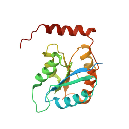
Ribose-5-phosphate isomerase is an enzyme that catalyzes the interconversion of ribose-5-phosphate and ribulose-5-phosphate. This family of enzymes naturally occurs in two distinct classes, RpiA and RpiB, which play an important role in the pentose phosphate pathway and nucleotide and co-factor biogenesis. Although RpiB occurs predominantly in bacteria, here we report crystal structures of a putative RpiB from the pathogenic fungus Coccidioides immitis. A 1.9 Å resolution apo structure was solved by combined molecular replacement and single wavelength anomalous dispersion (SAD) phasing using a crystal soaked briefly in a solution containing a high concentration of iodide ions. RpiB from C. immitis contains modest sequence and high structural homology to other known RpiB structures. A 1.8 Å resolution phosphate-bound structure demonstrates phosphate recognition and charge stabilization by a single positively charged residue whereas other members of this family use up to five positively charged residues to contact the phosphate of ribose-5-phosphate. A 1.7 Å resolution structure was obtained in which the catalytic base of C. immitis RpiB, Cys76, appears to form a weakly covalent bond with the central carbon of malonic acid with a bond distance of 2.2 Å. This interaction may mimic that formed by the suicide inhibitor iodoacetic acid with RpiB. The C. immitis RpiB contains the same fold and similar features as other members of this class of enzymes such as a highly reactive active site cysteine residue, but utilizes a divergent phosphate recognition strategy and may recognize a different substrate altogether.
- Seattle Structural Genomics Center for Infectious Disease (SSGCID), USA. tedwards@embios.com
Organizational Affiliation: