3SDZ | pdb_00003sdz

Structural characterization of the subunit A mutant F427W of the A-ATP synthase from Pyrococcus horikoshii


Experimental Data Snapshot

  • Method: X-RAY DIFFRACTION
  • Resolution: 2.53 Å
  • R-Value Free: 
    0.266 (Depositor), 0.271 (DCC) 
  • R-Value Work: 
    0.200 (Depositor), 0.205 (DCC) 
  • R-Value Observed: 
    0.203 (Depositor) 

Starting Model: experimental
View more details

wwPDB Validation 3D Report Full Report

Validation slider image for 3SDZ

This is version 1.2 of the entry. See complete history

Literature

Engineered tryptophan in the adenine-binding pocket of catalytic subunit A of A-ATP synthase demonstrates the importance of aromatic residues in adenine binding, forming a tool for steady-state and time-resolved fluorescence spectroscopy.

Tadwal, V.S.Manimekalai, M.S.Gruber, G.

(2011) Acta Crystallogr Sect F Struct Biol Cryst Commun 67: 1485-1491

  • DOI: https://doi.org/10.1107/S1744309111039595
  • Primary Citation Related Structures: 
    3SDZ, 3SE0

  • PubMed Abstract: 

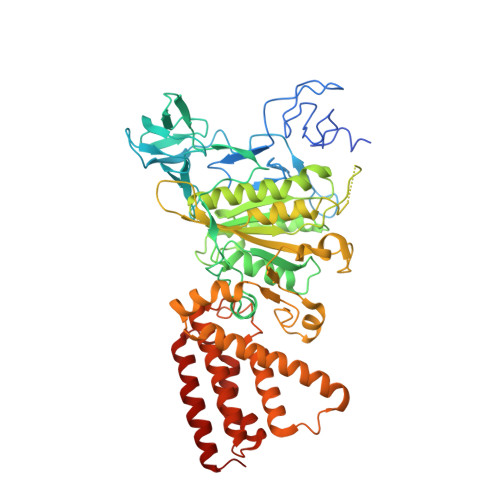
    A reporter tryptophan residue was individually introduced by site-directed mutagenesis into the adenine-binding pocket of the catalytic subunit A (F427W and F508W mutants) of the motor protein A(1)A(O) ATP synthase from Pyrococcus horikoshii OT3. The crystal structures of the F427W and F508W mutant proteins were determined to 2.5 and 2.6 Å resolution, respectively. The tryptophan substitution caused the fluorescence signal to increase by 28% (F427W) and 33% (F508W), with a shift from 333 nm in the wild-type protein to 339 nm in the mutant proteins. Tryptophan emission spectra showed binding of Mg-ATP to the F427W mutant with a K(d) of 8.5 µM. In contrast, no significant binding of nucleotide could be observed for the F508W mutant. A closer inspection of the crystal structure of the F427W mutant showed that the adenine-binding pocket had widened by 0.7 Å (to 8.70 Å) in comparison to the wild-type subunit A (8.07 Å) owing to tryptophan substitution, as a result of which it was able to bind ATP. In contrast, the adenine-binding pocket had narrowed in the F508W mutant. The two mutants presented demonstrate that the exact volume of the adenine ribose binding pocket is essential for nucleotide binding and even minor narrowing makes it unfit for nucleotide binding. In addition, structural and fluorescence data confirmed the viability of the fluorescently active mutant F427W, which had ideal tryptophan spectra for future structure-based time-resolved dynamic measurements of the catalytic subunit A of the ATP-synthesizing enzyme A-ATP synthase.


  • Organizational Affiliation
    • School of Biological Sciences, Nanyang Technological University, Singapore, Singapore.

Macromolecule Content 

  • Total Structure Weight: 67.15 kDa 
  • Atom Count: 4,701 
  • Modeled Residue Count: 548 
  • Deposited Residue Count: 588 
  • Unique protein chains: 1

Macromolecules

Find similar proteins by:|  3D Structure
Entity ID: 1
MoleculeChains  Sequence LengthOrganismDetailsImage
V-type ATP synthase alpha chain588Pyrococcus horikoshii OT3Mutation(s): 1 
Gene Names: atpAPH1975
EC: 3.6.3.14 (PDB Primary Data), 7.1.2.2 (UniProt)
UniProt
Find proteins for O57728 (Pyrococcus horikoshii (strain ATCC 700860 / DSM 12428 / JCM 9974 / NBRC 100139 / OT-3))
Explore O57728 
Go to UniProtKB:  O57728
Entity Groups
Sequence Clusters30% Identity50% Identity70% Identity90% Identity95% Identity100% Identity
UniProt GroupO57728
Sequence Annotations
Expand
Reference Sequence

Small Molecules

Ligands 3 Unique
IDChains Name / Formula / InChI Key2D Diagram3D Interactions
TRS

Query on TRS


Download:Ideal Coordinates CCD File
L [auth A],
M [auth A],
N [auth A]
2-AMINO-2-HYDROXYMETHYL-PROPANE-1,3-DIOL
C4 H12 N O3
LENZDBCJOHFCAS-UHFFFAOYSA-O
MPD

Query on MPD


Download:Ideal Coordinates CCD File
B [auth A]
C [auth A]
D [auth A]
E [auth A]
G [auth A]
B [auth A],
C [auth A],
D [auth A],
E [auth A],
G [auth A],
O [auth A]
(4S)-2-METHYL-2,4-PENTANEDIOL
C6 H14 O2
SVTBMSDMJJWYQN-YFKPBYRVSA-N
ACY

Query on ACY


Download:Ideal Coordinates CCD File
F [auth A]
H [auth A]
I [auth A]
J [auth A]
K [auth A]
F [auth A],
H [auth A],
I [auth A],
J [auth A],
K [auth A],
P [auth A]
ACETIC ACID
C2 H4 O2
QTBSBXVTEAMEQO-UHFFFAOYSA-N

Experimental Data & Validation

Experimental Data

  • Method: X-RAY DIFFRACTION
  • Resolution: 2.53 Å
  • R-Value Free:  0.266 (Depositor), 0.271 (DCC) 
  • R-Value Work:  0.200 (Depositor), 0.205 (DCC) 
  • R-Value Observed: 0.203 (Depositor) 
Space Group: P 43 21 2
Unit Cell:
Length ( Å )Angle ( ˚ )
a = 127.879α = 90
b = 127.879β = 90
c = 105.505γ = 90
Software Package:
Software NamePurpose
Blu-Icedata collection
MOLREPphasing
REFMACrefinement
HKL-2000data reduction
HKL-2000data scaling

Structure Validation

View Full Validation Report


Entry History 

Deposition Data

Revision History  (Full details and data files)

  • Version 1.0: 2012-01-25
    Type: Initial release
  • Version 1.1: 2017-08-23
    Changes: Refinement description, Source and taxonomy
  • Version 1.2: 2023-11-01
    Changes: Data collection, Database references, Derived calculations, Refinement description