3SBL | pdb_00003sbl

Crystal Structure of New Delhi Metal-beta-lactamase-1 from Klebsiella pneumoniae


Experimental Data Snapshot

  • Method: X-RAY DIFFRACTION
  • Resolution: 2.31 Å
  • R-Value Free: 
    0.260 (Depositor) 
  • R-Value Work: 
    0.200 (Depositor) 
  • R-Value Observed: 
    0.203 (Depositor) 

Starting Model: experimental
View more details

wwPDB Validation 3D Report Full Report

Validation slider image for 3SBL

This is version 1.4 of the entry. See complete history

Literature

Structure of Apo- and Monometalated Forms of NDM-1 A Highly Potent Carbapenem-Hydrolyzing Metallo-beta-Lactamase

Kim, Y.Tesar, C.Mire, J.Jedrzejczak, R.Binkowski, A.Babnigg, G.Sacchettini, J.Joachimiak, A.

(2011) PLoS One 6: e24621-e24621

  • DOI: https://doi.org/10.1371/journal.pone.0024621
  • Primary Citation Related Structures: 
    3RKJ, 3RKK, 3SBL, 3SFP

  • PubMed Abstract: 

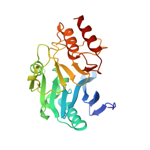
    The New Delhi Metallo-β-lactamase (NDM-1) gene makes multiple pathogenic microorganisms resistant to all known β-lactam antibiotics. The rapid emergence of NDM-1 has been linked to mobile plasmids that move between different strains resulting in world-wide dissemination. Biochemical studies revealed that NDM-1 is capable of efficiently hydrolyzing a wide range of β-lactams, including many carbapenems considered as "last resort" antibiotics. The crystal structures of metal-free apo- and monozinc forms of NDM-1 presented here revealed an enlarged and flexible active site of class B1 metallo-β-lactamase. This site is capable of accommodating many β-lactam substrates by having many of the catalytic residues on flexible loops, which explains the observed extended spectrum activity of this zinc dependent β-lactamase. Indeed, five loops contribute "keg" residues in the active site including side chains involved in metal binding. Loop 1 in particular, shows conformational flexibility, apparently related to the acceptance and positioning of substrates for cleavage by a zinc-activated water molecule.


  • Organizational Affiliation
    • Midwest Center for Structural Genomics and Structural Biology Center, Biosciences, Argonne National Laboratory, Argonne, Illinois, United States of America.

Macromolecule Content 

  • Total Structure Weight: 25.29 kDa 
  • Atom Count: 1,818 
  • Modeled Residue Count: 232 
  • Deposited Residue Count: 237 
  • Unique protein chains: 1

Macromolecules

Find similar proteins by:|  3D Structure
Entity ID: 1
MoleculeChains  Sequence LengthOrganismDetailsImage
Beta-lactamase NDM-1237Klebsiella pneumoniaeMutation(s): 0 
Gene Names: blaNDM-1
EC: 3.5.2.6
UniProt
Find proteins for C7C422 (Klebsiella pneumoniae)
Explore C7C422 
Go to UniProtKB:  C7C422
Entity Groups
Sequence Clusters30% Identity50% Identity70% Identity90% Identity95% Identity100% Identity
UniProt GroupC7C422
Sequence Annotations
Expand
Reference Sequence

Small Molecules

Ligands 1 Unique
IDChains Name / Formula / InChI Key2D Diagram3D Interactions
CIT

Query on CIT


Download:Ideal Coordinates CCD File
B [auth A]CITRIC ACID
C6 H8 O7
KRKNYBCHXYNGOX-UHFFFAOYSA-N

Experimental Data & Validation

Experimental Data

  • Method: X-RAY DIFFRACTION
  • Resolution: 2.31 Å
  • R-Value Free:  0.260 (Depositor) 
  • R-Value Work:  0.200 (Depositor) 
  • R-Value Observed: 0.203 (Depositor) 
Space Group: I 2 2 2
Unit Cell:
Length ( Å )Angle ( ˚ )
a = 66.05α = 90
b = 83.269β = 90
c = 105.401γ = 90
Software Package:
Software NamePurpose
SBC-Collectdata collection
HKL-3000data collection
HKL-3000phasing
MOLREPphasing
PHENIXrefinement
HKL-3000data reduction
HKL-3000data scaling

Structure Validation

View Full Validation Report


Entry History 

Revision History  (Full details and data files)

  • Version 1.0: 2011-06-29
    Type: Initial release
  • Version 1.1: 2011-07-13
    Changes: Version format compliance
  • Version 1.2: 2011-09-21
    Changes: Database references
  • Version 1.3: 2012-03-28
    Changes: Database references
  • Version 1.4: 2023-09-13
    Changes: Data collection, Database references, Derived calculations, Refinement description