3SAX | pdb_00003sax

Crystal structure of human carbonic anhydrase isozyme II with 2-chloro-5-{[(5-ethyl-2-pyrimidinyl)sulfanyl]acetyl}benzenesulfonamide


Experimental Data Snapshot

  • Method: X-RAY DIFFRACTION
  • Resolution: 1.10 Å
  • R-Value Free: 
    0.158 (Depositor), 0.162 (DCC) 
  • R-Value Work: 
    0.126 (Depositor), 0.133 (DCC) 
  • R-Value Observed: 
    0.129 (Depositor) 

Starting Model: experimental
View more details

wwPDB Validation 3D Report Full Report

Validation slider image for 3SAX

Ligand Structure Quality Assessment 


This is version 1.3 of the entry. See complete history

Literature

Design of [(2-pyrimidinylthio)acetyl]benzenesulfonamides as inhibitors of human carbonic anhydrases.

Capkauskaite, E.Zubriene, A.Baranauskiene, L.Tamulaitiene, G.Manakova, E.Kairys, V.Grazulis, S.Tumkevicius, S.Matulis, D.

(2012) Eur J Med Chem 51: 259-270

  • DOI: https://doi.org/10.1016/j.ejmech.2012.02.050
  • Primary Citation Related Structures: 
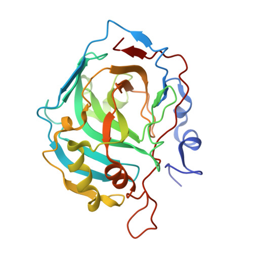
    3M2N, 3S8X, 3S9T, 3SAP, 3SAX, 3SBH, 3SBI

  • PubMed Abstract: 

    A series of [(2-pyrimidinylthio)acetyl]benzenesulfonamides were designed and synthesized. Their binding affinities as inhibitors of several recombinant human carbonic anhydrase (CA) isozymes were determined by isothermal titration calorimetry (ITC) and thermal shift assay (TSA). A group of compounds containing a chlorine atom in the benzenesulfonamide ring were found to exhibit higher selectivity but lower binding affinity toward tested CAs. The crystal structures of selected compounds in complex with CA II were determined to atomic resolution. Docking studies were performed to compare the binding modes of experimentally determined crystallographic structures with computational prediction of the pyrimidine derivative binding to CA II. Several compounds bound to select CAs with single-digit nanomolar affinities and could be used as leads for inhibitor development toward a select CA isozyme.


  • Organizational Affiliation
    • Vilnius University, Institute of Biotechnology, Department of Biothermodynamics and Drug Design, Graičiūno 8, Vilnius LT-02241, Lithuania.

Macromolecule Content 

  • Total Structure Weight: 30.05 kDa 
  • Atom Count: 3,190 
  • Modeled Residue Count: 258 
  • Deposited Residue Count: 260 
  • Unique protein chains: 1

Macromolecules

Find similar proteins by:|  3D Structure
Entity ID: 1
MoleculeChains  Sequence LengthOrganismDetailsImage
Carbonic anhydrase 2260Homo sapiensMutation(s): 0 
Gene Names: CA2
EC: 4.2.1.1 (PDB Primary Data), 4.2.1.69 (UniProt)
UniProt & NIH Common Fund Data Resources
Find proteins for P00918 (Homo sapiens)
Explore P00918 
Go to UniProtKB:  P00918
PHAROS:  P00918
GTEx:  ENSG00000104267 
Entity Groups
Sequence Clusters30% Identity50% Identity70% Identity90% Identity95% Identity100% Identity
UniProt GroupP00918
Sequence Annotations
Expand
Reference Sequence

Small Molecules

Ligands 4 Unique
IDChains Name / Formula / InChI Key2D Diagram3D Interactions
E50

Query on E50


Download:Ideal Coordinates CCD File
C [auth A]2-chloro-5-{[(5-ethylpyrimidin-2-yl)sulfanyl]acetyl}benzenesulfonamide
C14 H14 Cl N3 O3 S2
SAVHXXXMJJLUJH-UHFFFAOYSA-N
BCN

Query on BCN


Download:Ideal Coordinates CCD File
D [auth A]BICINE
C6 H13 N O4
FSVCELGFZIQNCK-UHFFFAOYSA-N
DMS

Query on DMS


Download:Ideal Coordinates CCD File
E [auth A],
F [auth A]
DIMETHYL SULFOXIDE
C2 H6 O S
IAZDPXIOMUYVGZ-UHFFFAOYSA-N
ZN

Query on ZN


Download:Ideal Coordinates CCD File
B [auth A]ZINC ION
Zn
PTFCDOFLOPIGGS-UHFFFAOYSA-N
Binding Affinity Annotations 
IDSourceBinding Affinity
E50 BindingDB:  3SAX Kd: min: 140, max: 270 (nM) from 3 assay(s)

Experimental Data & Validation

Experimental Data

  • Method: X-RAY DIFFRACTION
  • Resolution: 1.10 Å
  • R-Value Free:  0.158 (Depositor), 0.162 (DCC) 
  • R-Value Work:  0.126 (Depositor), 0.133 (DCC) 
  • R-Value Observed: 0.129 (Depositor) 
Space Group: P 1 21 1
Unit Cell:
Length ( Å )Angle ( ˚ )
a = 42.221α = 90
b = 41.172β = 104.2
c = 72.197γ = 90
Software Package:
Software NamePurpose
REFMACrefinement
MOSFLMdata reduction
SCALAdata scaling
PDB_EXTRACTdata extraction
DNAdata collection

Structure Validation

View Full Validation Report


Ligand Structure Quality Assessment 


Entry History 

Deposition Data

Revision History  (Full details and data files)

  • Version 1.0: 2012-04-11
    Type: Initial release
  • Version 1.1: 2012-05-16
    Changes: Database references
  • Version 1.2: 2017-11-15
    Changes: Database references
  • Version 1.3: 2023-09-13
    Changes: Data collection, Database references, Derived calculations, Refinement description