3SAS

MUTM Slanted complex 4 with R112A mutation


Experimental Data Snapshot

  • Method: X-RAY DIFFRACTION
  • Resolution: 2.05 Å
  • R-Value Free: 0.213 
  • R-Value Work: 0.180 
  • R-Value Observed: 0.182 

wwPDB Validation   3D Report Full Report


This is version 1.4 of the entry. See complete history


Literature

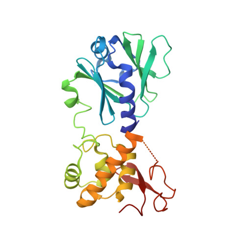
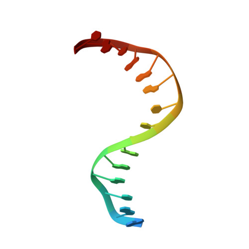
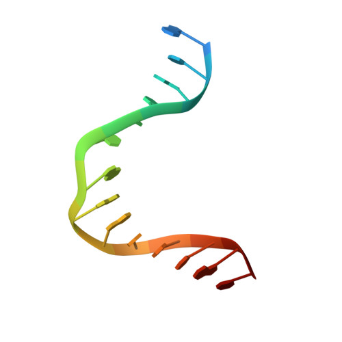
Strandwise translocation of a DNA glycosylase on undamaged DNA.

Qi, Y.Nam, K.Spong, M.C.Banerjee, A.Sung, R.J.Zhang, M.Karplus, M.Verdine, G.L.

(2012) Proc Natl Acad Sci U S A 109: 1086-1091

  • DOI: https://doi.org/10.1073/pnas.1111237108
  • Primary Citation of Related Structures:  
    3SAR, 3SAS, 3SAT, 3SAU, 3SAV, 3SAW, 3SBJ

  • PubMed Abstract: 

    Base excision repair of genotoxic nucleobase lesions in the genome is critically dependent upon the ability of DNA glycosylases to locate rare sites of damage embedded in a vast excess of undamaged DNA, using only thermal energy to fuel the search process. Considerable interest surrounds the question of how DNA glycosylases translocate efficiently along DNA while maintaining their vigilance for target damaged sites. Here, we report the observation of strandwise translocation of 8-oxoguanine DNA glycosylase, MutM, along undamaged DNA. In these complexes, the protein is observed to translocate by one nucleotide on one strand while remaining untranslocated on the complementary strand. We further report that alterations of single base-pairs or a single amino acid substitution (R112A) can induce strandwise translocation. Molecular dynamics simulations confirm that MutM can translocate along DNA in a strandwise fashion. These observations reveal a previously unobserved mode of movement for a DNA-binding protein along the surface of DNA.


  • Organizational Affiliation

    Program in Biophysics, Harvard Medical School, 240 Longwood Avenue, Boston, MA 02115, USA.


Macromolecules

Find similar proteins by:  (by identity cutoff)  |  3D Structure
Entity ID: 1
MoleculeChains Sequence LengthOrganismDetailsImage
DNA GLYCOSYLASE273Geobacillus stearothermophilusMutation(s): 2 
Gene Names: MUTM
EC: 4.2.99.18
UniProt
Find proteins for P84131 (Geobacillus stearothermophilus)
Explore P84131 
Go to UniProtKB:  P84131
Entity Groups  
Sequence Clusters30% Identity50% Identity70% Identity90% Identity95% Identity100% Identity
UniProt GroupP84131
Sequence Annotations
Expand
  • Reference Sequence

Find similar nucleic acids by:  Sequence   |   3D Structure  

Entity ID: 2
MoleculeChains LengthOrganismImage
5'-D(*A*GP*GP*TP*AP*GP*AP*CP*TP*CP*GP*GP*AP*CP*GP*C)-3'16N/A
Sequence Annotations
Expand
  • Reference Sequence

Find similar nucleic acids by:  Sequence   |   3D Structure  

Entity ID: 3
MoleculeChains LengthOrganismImage
5'-D(*T*GP*CP*GP*TP*CP*CP*GP*AP*GP*(TX2) P*CP*TP*AP*CP*C)-3'16N/A
Sequence Annotations
Expand
  • Reference Sequence
Small Molecules
Ligands 1 Unique
IDChains Name / Formula / InChI Key2D Diagram3D Interactions
ZN
Query on ZN

Download Ideal Coordinates CCD File 
D [auth A]ZINC ION
Zn
PTFCDOFLOPIGGS-UHFFFAOYSA-N
Experimental Data & Validation

Experimental Data

  • Method: X-RAY DIFFRACTION
  • Resolution: 2.05 Å
  • R-Value Free: 0.213 
  • R-Value Work: 0.180 
  • R-Value Observed: 0.182 
  • Space Group: P 21 21 21
Unit Cell:
Length ( Å )Angle ( ˚ )
a = 45.393α = 90
b = 96.029β = 90
c = 103.369γ = 90
Software Package:
Software NamePurpose
PHENIXrefinement
CNSrefinement
DENZOdata reduction
SCALEPACKdata scaling
CNSphasing

Structure Validation

View Full Validation Report


Entry History 

Deposition Data

Revision History  (Full details and data files)

  • Version 1.0: 2012-01-11
    Type: Initial release
  • Version 1.1: 2012-02-22
    Changes: Database references
  • Version 1.2: 2017-11-08
    Changes: Refinement description
  • Version 1.3: 2019-07-17
    Changes: Data collection, Derived calculations, Refinement description
  • Version 1.4: 2023-09-13
    Changes: Data collection, Database references, Derived calculations, Refinement description