Structural insights into apoptotic DNA degradation by CED-3 protease suppressor-6 (CPS-6) from Caenorhabditis elegans
Lin, J.L.J., Nakagawa, A., Lin, C.L., Hsiao, Y.Y., Yang, W.Z., Wang, Y.T., Doudeva, L.G., Skeen-Gaar, R.R., Xue, D., Yuan, H.S.(2012) J Biological Chem 287: 7110-7120
- PubMed: 22223640 Search on PubMedSearch on PubMed Central
- DOI: https://doi.org/10.1074/jbc.M111.316075
- Primary Citation Related Structures:
3S5B - PubMed Abstract:
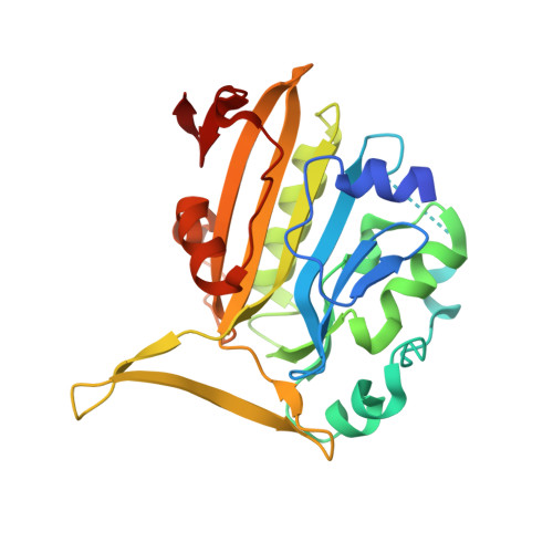
Endonuclease G (EndoG) is a mitochondrial protein that traverses to the nucleus and participates in chromosomal DNA degradation during apoptosis in yeast, worms, flies, and mammals. However, it remains unclear how EndoG binds and digests DNA. Here we show that the Caenorhabditis elegans CPS-6, a homolog of EndoG, is a homodimeric Mg(2+)-dependent nuclease, binding preferentially to G-tract DNA in the optimum low salt buffer at pH 7. The crystal structure of CPS-6 was determined at 1.8 Å resolution, revealing a mixed αβ topology with the two ββα-metal finger nuclease motifs located distantly at the two sides of the dimeric enzyme. A structural model of the CPS-6-DNA complex suggested a positively charged DNA-binding groove near the Mg(2+)-bound active site. Mutations of four aromatic and basic residues: Phe(122), Arg(146), Arg(156), and Phe(166), in the protein-DNA interface significantly reduced the DNA binding and cleavage activity of CPS-6, confirming that these residues are critical for CPS-6-DNA interactions. In vivo transformation rescue experiments further showed that the reduced DNase activity of CPS-6 mutants was positively correlated with its diminished cell killing activity in C. elegans. Taken together, these biochemical, structural, mutagenesis, and in vivo data reveal a molecular basis of how CPS-6 binds and hydrolyzes DNA to promote cell death.
- Institute of Molecular Biology, Academia Sinica, Taipei 11529, Taiwan.
Organizational Affiliation: