3RZW

Crystal Structure of the Monobody ySMB-9 bound to human SUMO1


Experimental Data Snapshot

  • Method: X-RAY DIFFRACTION
  • Resolution: 2.15 Å
  • R-Value Free: 0.237 
  • R-Value Work: 0.186 
  • R-Value Observed: 0.189 

wwPDB Validation   3D Report Full Report


This is version 1.3 of the entry. See complete history


Literature

Teaching an old scaffold new tricks: monobodies constructed using alternative surfaces of the FN3 scaffold.

Koide, A.Wojcik, J.Gilbreth, R.N.Hoey, R.J.Koide, S.

(2012) J Mol Biol 415: 393-405

  • DOI: https://doi.org/10.1016/j.jmb.2011.12.019
  • Primary Citation of Related Structures:  
    3RZW, 3UYO

  • PubMed Abstract: 

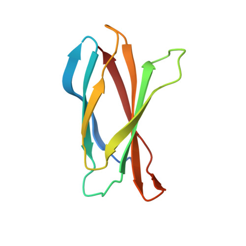
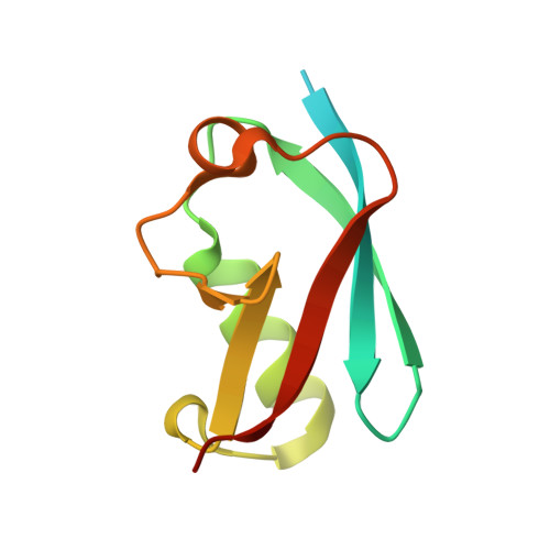
    The fibronectin type III domain (FN3) has become one of the most widely used non-antibody scaffolds for generating new binding proteins. Because of its structural homology to the immunoglobulin domain, combinatorial libraries of FN3 designed to date have primarily focused on introducing amino acid diversity into three loops that are equivalent to antibody complementarity-determining regions. Here, we report an FN3 library that utilizes alternative positions for presenting amino acid diversity. We diversified positions on a β-sheet and surface loops that together form a concave surface. The new library produced binding proteins (termed "monobodies") to multiple target proteins, generally with similar efficacy as the original, loop-focused library. The crystal structure of a monobody generated from the new library in complex with its target, the Abl SH2 domain, revealed that a concave surface of the monobody, as intended in our design, bound to a convex surface of the target with the interface area being among the largest of published structures of monobody-target complexes. This mode of interaction differs from a common binding mode for single-domain antibodies and antibody mimics in which recognition loops recognize clefts in targets. Together, this work illustrates the utilization of different surfaces of a single immunoglobulin-like scaffold to generate binding proteins with distinct characteristics.


  • Organizational Affiliation

    Department of Biochemistry and Molecular Biology, The University of Chicago, 929 East 57th Street, Chicago,IL 60637, USA.


Macromolecules
Find similar proteins by:  (by identity cutoff)  |  3D Structure
Entity ID: 1
MoleculeChains Sequence LengthOrganismDetailsImage
Monobody ySMB-9
A, B
95Homo sapiensMutation(s): 0 
Gene Names: SUMO1SMT3CSMT3H3UBL1OK/SW-cl.43
Entity Groups  
Sequence Clusters30% Identity50% Identity70% Identity90% Identity95% Identity100% Identity
Sequence Annotations
Expand
  • Reference Sequence
Find similar proteins by:  (by identity cutoff)  |  3D Structure
Entity ID: 2
MoleculeChains Sequence LengthOrganismDetailsImage
Small ubiquitin-related modifier 1
C, D
99Homo sapiensMutation(s): 1 
UniProt & NIH Common Fund Data Resources
Find proteins for P63165 (Homo sapiens)
Explore P63165 
Go to UniProtKB:  P63165
PHAROS:  P63165
GTEx:  ENSG00000116030 
Entity Groups  
Sequence Clusters30% Identity50% Identity70% Identity90% Identity95% Identity100% Identity
UniProt GroupP63165
Sequence Annotations
Expand
  • Reference Sequence
Experimental Data & Validation

Experimental Data

  • Method: X-RAY DIFFRACTION
  • Resolution: 2.15 Å
  • R-Value Free: 0.237 
  • R-Value Work: 0.186 
  • R-Value Observed: 0.189 
  • Space Group: C 2 2 21
Unit Cell:
Length ( Å )Angle ( ˚ )
a = 93.347α = 90
b = 97.829β = 90
c = 96.578γ = 90
Software Package:
Software NamePurpose
MAR345data collection
MD2data collection
MOLREPphasing
REFMACrefinement
HKL-2000data reduction
HKL-2000data scaling

Structure Validation

View Full Validation Report


Entry History 

Deposition Data

Revision History  (Full details and data files)

  • Version 1.0: 2011-12-28
    Type: Initial release
  • Version 1.1: 2012-05-23
    Changes: Database references
  • Version 1.2: 2017-11-08
    Changes: Refinement description
  • Version 1.3: 2024-02-28
    Changes: Data collection, Database references, Derived calculations