3RNM

The crystal structure of the subunit binding of human dihydrolipoamide transacylase (E2b) bound to human dihydrolipoamide dehydrogenase (E3)


Experimental Data Snapshot

  • Method: X-RAY DIFFRACTION
  • Resolution: 2.40 Å
  • R-Value Free: 0.239 
  • R-Value Work: 0.181 
  • R-Value Observed: 0.184 

wwPDB Validation   3D Report Full Report


Ligand Structure Quality Assessment 


This is version 1.2 of the entry. See complete history


Literature

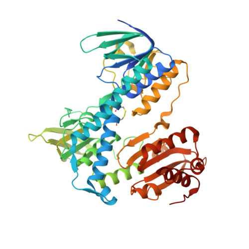
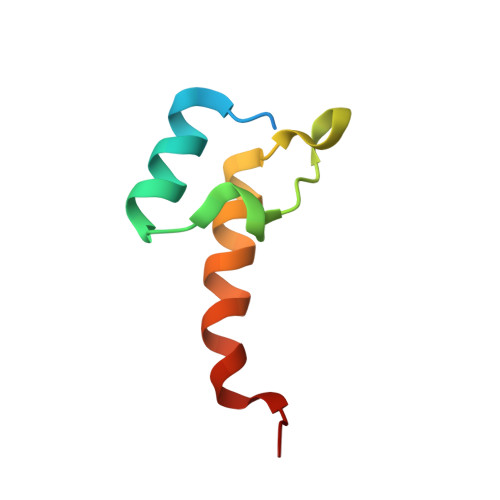
Structural and Thermodynamic Basis for Weak Interactions between Dihydrolipoamide Dehydrogenase and Subunit-binding Domain of the Branched-chain {alpha}-Ketoacid Dehydrogenase Complex.

Brautigam, C.A.Wynn, R.M.Chuang, J.L.Naik, M.T.Young, B.B.Huang, T.H.Chuang, D.T.

(2011) J Biol Chem 286: 23476-23488

  • DOI: https://doi.org/10.1074/jbc.M110.202960
  • Primary Citation of Related Structures:  
    3RNM

  • PubMed Abstract: 

    The purified mammalian branched-chain α-ketoacid dehydrogenase complex (BCKDC), which catalyzes the oxidative decarboxylation of branched-chain α-keto acids, is essentially devoid of the constituent dihydrolipoamide dehydrogenase component (E3). The absence of E3 is associated with the low affinity of the subunit-binding domain of human BCKDC (hSBDb) for hE3. In this work, sequence alignments of hSBDb with the E3-binding domain (E3BD) of the mammalian pyruvate dehydrogenase complex show that hSBDb has an arginine at position 118, where E3BD features an asparagine. Substitution of Arg-118 with an asparagine increases the binding affinity of the R118N hSBDb variant (designated hSBDb*) for hE3 by nearly 2 orders of magnitude. The enthalpy of the binding reaction changes from endothermic with the wild-type hSBDb to exothermic with the hSBDb* variant. This higher affinity interaction allowed the determination of the crystal structure of the hE3/hSBDb* complex to 2.4-Å resolution. The structure showed that the presence of Arg-118 poses a unique, possibly steric and/or electrostatic incompatibility that could impede E3 interactions with the wild-type hSBDb. Compared with the E3/E3BD structure, the hE3/hSBDb* structure has a smaller interfacial area. Solution NMR data corroborated the interactions of hE3 with Arg-118 and Asn-118 in wild-type hSBDb and mutant hSBDb*, respectively. The NMR results also showed that the interface between hSBDb and hE3 does not change significantly from hSBDb to hSBDb*. Taken together, our results represent a starting point for explaining the long standing enigma that the E2b core of the BCKDC binds E3 far more weakly relative to other α-ketoacid dehydrogenase complexes.


  • Organizational Affiliation

    Department of Biochemistry, University of Texas Southwestern Medical Center, Dallas, Texas 75390, USA. chad.brautigam@utsouthwestern.edu


Macromolecules
Find similar proteins by:  (by identity cutoff)  |  3D Structure
Entity ID: 1
MoleculeChains Sequence LengthOrganismDetailsImage
Dihydrolipoyl dehydrogenase, mitochondrial
A, B, C, D
495Homo sapiensMutation(s): 0 
Gene Names: DLDGCSLLADPHE3
EC: 1.8.1.4
UniProt & NIH Common Fund Data Resources
Find proteins for P09622 (Homo sapiens)
Explore P09622 
Go to UniProtKB:  P09622
PHAROS:  P09622
GTEx:  ENSG00000091140 
Entity Groups  
Sequence Clusters30% Identity50% Identity70% Identity90% Identity95% Identity100% Identity
UniProt GroupP09622
Sequence Annotations
Expand
  • Reference Sequence
Find similar proteins by:  (by identity cutoff)  |  3D Structure
Entity ID: 2
MoleculeChains Sequence LengthOrganismDetailsImage
Lipoamide acyltransferase component of branched-chain alpha-keto acid dehydrogenase complex, mitochondrial
E, F
58Homo sapiensMutation(s): 1 
Gene Names: DBTBCATE2
EC: 2.3.1.168
UniProt & NIH Common Fund Data Resources
Find proteins for P11182 (Homo sapiens)
Explore P11182 
Go to UniProtKB:  P11182
PHAROS:  P11182
GTEx:  ENSG00000137992 
Entity Groups  
Sequence Clusters30% Identity50% Identity70% Identity90% Identity95% Identity100% Identity
UniProt GroupP11182
Sequence Annotations
Expand
  • Reference Sequence
Small Molecules
Ligands 3 Unique
IDChains Name / Formula / InChI Key2D Diagram3D Interactions
FAD
Query on FAD

Download Ideal Coordinates CCD File 
H [auth A],
K [auth B],
N [auth C],
Q [auth D]
FLAVIN-ADENINE DINUCLEOTIDE
C27 H33 N9 O15 P2
VWWQXMAJTJZDQX-UYBVJOGSSA-N
NHE
Query on NHE

Download Ideal Coordinates CCD File 
I [auth A],
L [auth B],
O [auth C],
R [auth D]
2-[N-CYCLOHEXYLAMINO]ETHANE SULFONIC ACID
C8 H17 N O3 S
MKWKNSIESPFAQN-UHFFFAOYSA-N
BME
Query on BME

Download Ideal Coordinates CCD File 
G [auth A],
J [auth B],
M [auth C],
P [auth D]
BETA-MERCAPTOETHANOL
C2 H6 O S
DGVVWUTYPXICAM-UHFFFAOYSA-N
Experimental Data & Validation

Experimental Data

  • Method: X-RAY DIFFRACTION
  • Resolution: 2.40 Å
  • R-Value Free: 0.239 
  • R-Value Work: 0.181 
  • R-Value Observed: 0.184 
  • Space Group: P 1 21 1
Unit Cell:
Length ( Å )Angle ( ˚ )
a = 84.136α = 90
b = 112.308β = 91.4
c = 122.545γ = 90
Software Package:
Software NamePurpose
SBC-Collectdata collection
PHENIXmodel building
PHENIXrefinement
HKL-3000data reduction
HKL-3000data scaling
PHENIXphasing

Structure Validation

View Full Validation Report


Ligand Structure Quality Assessment 


Entry History 

Deposition Data

Revision History  (Full details and data files)

  • Version 1.0: 2011-05-04
    Type: Initial release
  • Version 1.1: 2011-07-13
    Changes: Version format compliance
  • Version 1.2: 2011-07-20
    Changes: Database references